 Movie
Movie Controller


+ Open data

- Basic information
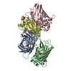
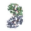
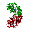
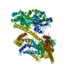
| Entry | Database: PDB / ID: 1b9c | |||||||||
|---|---|---|---|---|---|---|---|---|---|---|
| Title | Green Fluorescent Protein Mutant F99S, M153T and V163A | |||||||||
 Components Components | PROTEIN (GREEN FLUORESCENT PROTEIN) | |||||||||
 Keywords Keywords | LUMINESCENT PROTEIN / FLUORESCENT PROTEIN / CHROMOPHORE / GREEN FLUORESCENT PROTEIN / LUMINESCENCE / F99S M153T V163A MUTANT | |||||||||
| Function / homology |  Function and homology information Function and homology information | |||||||||
| Biological species |   Aequorea victoria (jellyfish) Aequorea victoria (jellyfish) | |||||||||
| Method |  X-RAY DIFFRACTION / SYNCHROTRON / MOLECULAR REPLACEMENT / Resolution: 2.4 Å X-RAY DIFFRACTION / SYNCHROTRON / MOLECULAR REPLACEMENT / Resolution: 2.4 Å | |||||||||
 Authors Authors | Battistutta, R. / Negro, A. / Zanotti, G. | |||||||||
 Citation Citation | Journal: Proteins / Year: 2000 Title: Crystal structure and refolding properties of the mutant F99S/M153T/V163A of the green fluorescent protein. Authors: Battistutta, R. / Negro, A. / Zanotti, G. #1: Journal: Nat.Biotechnol. / Year: 1996Title: The Molecular Structure of Green Fluorescent Protein Authors: Yang, F. / Moss, L.G. / Phillips Jr., G.N. #2: Journal: Science / Year: 1996Title: Crystal Structure of the Aequorea Victoria Green Fluorescent Protein Authors: Ormo, M. / Cubitt, A.B. / Kallio, K. / Gross, L.A. / Tsien, R.Y. / Remington, S.J. | |||||||||
| History |
|
- Structure visualization
| Structure viewer | Molecule: MolmilJmol/JSmol |
|---|
- Downloads & links
-Download
| PDBx/mmCIF format | 1b9c.cif.gz | 192.3 KB | Display | PDBx/mmCIF format |
|---|---|---|---|---|
| PDB format | pdb1b9c.ent.gz | 154 KB | Display | PDB format |
| PDBx/mmJSON format | 1b9c.json.gz | Tree view | PDBx/mmJSON format | |
| Others |  Other downloads Other downloads |
-Validation report
| Arichive directory | https://data.pdbj.org/pub/pdb/validation_reports/b9/1b9cftp://data.pdbj.org/pub/pdb/validation_reports/b9/1b9c | HTTPS FTP |
|---|
-Related structure data
| Related structure data | 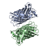 1gflS S: Starting model for refinement |
|---|---|
| Similar structure data |


-Links
 PDBj
PDBj

- Assembly
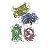
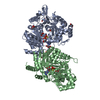
| Deposited unit | 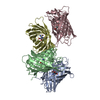
| ||||||||||||
|---|---|---|---|---|---|---|---|---|---|---|---|---|---|
| 1 |
| ||||||||||||
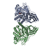
| Unit cell |
| ||||||||||||
| Noncrystallographic symmetry (NCS) | NCS oper:
|
-Components
| #1: Protein | Mass: 26737.959 Da / Num. of mol.: 4 / Mutation: F99S, M153T, V163A Source method: isolated from a genetically manipulated source Source: (gene. exp.) Aequorea victoria (jellyfish) / Description: THE N-TERMINAL HIS-TAG HAS BEEN REMOVED / Plasmid: PRSETB (INVITROGEN) / Species (production host): Escherichia coli / Production host:  #2: Water | ChemComp-HOH / |  Mass: 18.015 Da / Num. of mol.: 286 / Source method: isolated from a natural source / Formula: H2O Mass: 18.015 Da / Num. of mol.: 286 / Source method: isolated from a natural source / Formula: H2OHas protein modification | Y | Sequence details | THE FLUOROPHORE IS FORMED BY SER 65, TYR 66 AND GLY 67. THE CARBONYL CARBON OF TYR 66 IS BONDED TO ...THE FLUOROPHOR | |
|---|
-Experimental details
-Experiment
| Experiment | Method: X-RAY DIFFRACTION / Number of used crystals: 2 |
|---|
- Sample preparation
| Crystal | Density Matthews: 2.3 Å3/Da / Density % sol: 50 % | ||||||||||||||||||||||||||||||||||||||||||
|---|---|---|---|---|---|---|---|---|---|---|---|---|---|---|---|---|---|---|---|---|---|---|---|---|---|---|---|---|---|---|---|---|---|---|---|---|---|---|---|---|---|---|---|
| Crystal grow | pH: 8.3 Details: 22% PEG 4000, 50 MM HEPES PH 8.5, 50 MM MGCL2, 10 MM 2-MERCAPTOETHANOL, 23% MG/ ML PROTEINA, pH 8.3 | ||||||||||||||||||||||||||||||||||||||||||
| Crystal grow | *PLUS Temperature: 20 ℃ / pH: 7.4 / Method: vapor diffusion, hanging drop | ||||||||||||||||||||||||||||||||||||||||||
| Components of the solutions | *PLUS
|
-Data collection
| Diffraction | Mean temperature: 100 K |
|---|---|
| Diffraction source | Source: SYNCHROTRON / Site: ELETTRA  / Beamline: 5.2R / Wavelength: 1 / Beamline: 5.2R / Wavelength: 1 |
| Detector | Type: MAR scanner 345 mm plate / Detector: IMAGE PLATE |
| Radiation | Monochromator: SI (111) / Protocol: SINGLE WAVELENGTH / Monochromatic (M) / Laue (L): M / Scattering type: x-ray |
| Radiation wavelength | Wavelength: 1 Å / Relative weight: 1 |
| Reflection | Resolution: 2.37→30 Å / Num. obs: 36746 / % possible obs: 83.9 % / Observed criterion σ(I): 0 / Redundancy: 4.6 % / Rmerge(I) obs: 0.062 / Net I/σ(I): 6.9 |
| Reflection shell | Resolution: 2.37→2.64 Å / Redundancy: 3.9 % / Rmerge(I) obs: 0.146 / Mean I/σ(I) obs: 4.2 / % possible all: 45 |
| Reflection | *PLUS Num. measured all: 165357 |
| Reflection shell | *PLUS % possible obs: 45 % / Num. unique obs: 5445 |
- Processing
| Software |
| ||||||||||||||||||||||||||||||||||||||||||||||||||||||||||||
|---|---|---|---|---|---|---|---|---|---|---|---|---|---|---|---|---|---|---|---|---|---|---|---|---|---|---|---|---|---|---|---|---|---|---|---|---|---|---|---|---|---|---|---|---|---|---|---|---|---|---|---|---|---|---|---|---|---|---|---|---|---|
| Refinement | Method to determine structure: MOLECULAR REPLACEMENT Starting model: PDB ENTRY 1GFL Resolution: 2.4→30 Å / Isotropic thermal model: RESTRAINED / Cross valid method: THROUGHOUT / σ(F): 2 / Details: APPLIED DATA CUTOFF: F(CALC)/F (OBS) < = 3.5
| ||||||||||||||||||||||||||||||||||||||||||||||||||||||||||||
| Refinement step | Cycle: LAST / Resolution: 2.4→30 Å
| ||||||||||||||||||||||||||||||||||||||||||||||||||||||||||||
| Refine LS restraints |
| ||||||||||||||||||||||||||||||||||||||||||||||||||||||||||||
| Refine LS restraints NCS | NCS model details: RESTRAINTS | ||||||||||||||||||||||||||||||||||||||||||||||||||||||||||||
| Software | *PLUS Name: X-PLOR / Version: 3.8.5 / Classification: refinement | ||||||||||||||||||||||||||||||||||||||||||||||||||||||||||||
| Refinement | *PLUS Highest resolution: 2.4 Å / Lowest resolution: 30 Å / σ(F): 2 / % reflection Rfree: 10 % / Rfactor Rfree: 0.28 | ||||||||||||||||||||||||||||||||||||||||||||||||||||||||||||
| Solvent computation | *PLUS | ||||||||||||||||||||||||||||||||||||||||||||||||||||||||||||
| Displacement parameters | *PLUS | ||||||||||||||||||||||||||||||||||||||||||||||||||||||||||||
| Refine LS restraints | *PLUS
|
