 Movie
Movie Controller

[English] 日本語
 Yorodumi
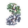
- PDB-6d99: Crystal structure of Corynebacterium diphtheriae UDP-galactopyran... -
+ Open data

- Basic information
| Entry | Database: PDB / ID: 6d99 | ||||||||||||
|---|---|---|---|---|---|---|---|---|---|---|---|---|---|
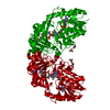
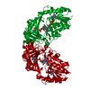
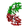
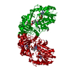
| Title | Crystal structure of Corynebacterium diphtheriae UDP-galactopyranose mutase in complex with UDP-GalNAc (open, oxidized) | ||||||||||||
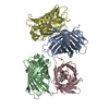
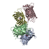
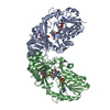
 Components Components | UDP-galactopyranose mutase | ||||||||||||
 Keywords Keywords | ISOMERASE / galatofuranose / galactopyranose / galactose / UDP / enzyme / UDP-galactopyranose mutase | ||||||||||||
| Function / homology | NAD(P)-binding Rossmann-like Domain / Rossmann fold / 3-Layer(aba) Sandwich / Alpha Beta / FLAVIN-ADENINE DINUCLEOTIDE / ISOPROPYL ALCOHOL / URIDINE-DIPHOSPHATE-N-ACETYLGALACTOSAMINE / :  Function and homology information Function and homology information | ||||||||||||
| Biological species |  Corynebacterium diphtheriae (bacteria) Corynebacterium diphtheriae (bacteria) | ||||||||||||
| Method |  X-RAY DIFFRACTION / SYNCHROTRON / MOLECULAR REPLACEMENT / molecular replacement / Resolution: 2.15 Å X-RAY DIFFRACTION / SYNCHROTRON / MOLECULAR REPLACEMENT / molecular replacement / Resolution: 2.15 Å | ||||||||||||
 Authors Authors | Wangkanont, K. / Kiessling, L.L. / Forest, K.T. | ||||||||||||
| Funding support |  United States, 3items United States, 3items
| ||||||||||||
 Citation Citation | Journal: To be published Title: Substrate recognition by FAD in UDP-galactopyranose mutase Authors: Wangkanont, K. / Forest, K.T. / Kiessling, L.L. | ||||||||||||
| History |
|
- Structure visualization
| Structure viewer | Molecule: MolmilJmol/JSmol |
|---|
- Downloads & links
-Download
| PDBx/mmCIF format | 6d99.cif.gz | 106.7 KB | Display | PDBx/mmCIF format |
|---|---|---|---|---|
| PDB format | pdb6d99.ent.gz | 77.5 KB | Display | PDB format |
| PDBx/mmJSON format | 6d99.json.gz | Tree view | PDBx/mmJSON format | |
| Others |  Other downloads Other downloads |
-Validation report
| Arichive directory | https://data.pdbj.org/pub/pdb/validation_reports/d9/6d99ftp://data.pdbj.org/pub/pdb/validation_reports/d9/6d99 | HTTPS FTP |
|---|
-Related structure data
| Related structure data |  6d2eC  6d2gC  6d9aC 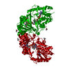 6d9bC  6d9cC 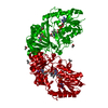 6d9dC  6d9eC 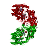 5br7S S: Starting model for refinement C: citing same article ( |
|---|---|
| Similar structure data |


-Links
 PDBj
PDBj
- Assembly
| Deposited unit | 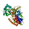
| ||||||||
|---|---|---|---|---|---|---|---|---|---|
| 1 | 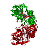
| ||||||||
| Unit cell |
|
-Components
-Protein , 1 types, 1 molecules A
| #1: Protein | Mass: 45737.668 Da / Num. of mol.: 1 / Fragment: UDP-galactopyranose mutase Source method: isolated from a genetically manipulated source Source: (gene. exp.) Corynebacterium diphtheriae (bacteria) / Gene: BU167_07490 / Plasmid: modified pMAL C5x / Production host: |
|---|
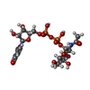
-Non-polymers , 5 types, 256 molecules 


| #2: Chemical | ChemComp-FAD /  Mass: 785.550 Da / Num. of mol.: 1 / Source method: obtained synthetically / Formula: C27H33N9O15P2 / Comment: FAD*YM Mass: 785.550 Da / Num. of mol.: 1 / Source method: obtained synthetically / Formula: C27H33N9O15P2 / Comment: FAD*YM | ||
|---|---|---|---|
| #3: Chemical | ChemComp-NA /  Mass: 22.990 Da / Num. of mol.: 1 / Source method: obtained synthetically / Formula: Na Mass: 22.990 Da / Num. of mol.: 1 / Source method: obtained synthetically / Formula: Na | ||
| #4: Chemical | ChemComp-UD2 /  Mass: 607.354 Da / Num. of mol.: 1 / Source method: obtained synthetically / Formula: C17H27N3O17P2 Mass: 607.354 Da / Num. of mol.: 1 / Source method: obtained synthetically / Formula: C17H27N3O17P2 | ||
| #5: Chemical | ChemComp-IPA /  Mass: 60.095 Da / Num. of mol.: 5 / Source method: obtained synthetically / Formula: C3H8O Mass: 60.095 Da / Num. of mol.: 5 / Source method: obtained synthetically / Formula: C3H8O#6: Water | ChemComp-HOH / | Mass: 18.015 Da / Num. of mol.: 248 / Source method: isolated from a natural source / Formula: H2O |
-Experimental details
-Experiment
| Experiment | Method: X-RAY DIFFRACTION / Number of used crystals: 1 |
|---|
- Sample preparation
| Crystal | Density Matthews: 3.34 Å3/Da / Density % sol: 63.19 % |
|---|---|
| Crystal grow | Temperature: 298 K / Method: vapor diffusion, hanging drop / pH: 8 / Details: 100 mM Tris, 28% PEG400, 20% isopropanol |
-Data collection
| Diffraction | Mean temperature: 100 K | ||||||||||||||||||||||||
|---|---|---|---|---|---|---|---|---|---|---|---|---|---|---|---|---|---|---|---|---|---|---|---|---|---|
| Diffraction source | Source: SYNCHROTRON / Site: APS / Beamline: 21-ID-F / Wavelength: 0.97872 Å | ||||||||||||||||||||||||
| Detector | Type: MARMOSAIC 225 mm CCD / Detector: CCD / Date: Mar 13, 2014 | ||||||||||||||||||||||||
| Radiation | Monochromator: C(111) / Protocol: SINGLE WAVELENGTH / Monochromatic (M) / Laue (L): M / Scattering type: x-ray | ||||||||||||||||||||||||
| Radiation wavelength | Wavelength: 0.97872 Å / Relative weight: 1 | ||||||||||||||||||||||||
| Reflection | Resolution: 2.15→46.87 Å / Num. obs: 64409 / % possible obs: 100 % / Redundancy: 29.1 % / Biso Wilson estimate: 36.23 Å2 / CC1/2: 0.998 / Rmerge(I) obs: 0.156 / Rpim(I) all: 0.029 / Rrim(I) all: 0.158 / Net I/σ(I): 14.4 | ||||||||||||||||||||||||
| Reflection shell | Diffraction-ID: 1
|
-Phasing
| Phasing | Method: molecular replacement | |||||||||
|---|---|---|---|---|---|---|---|---|---|---|
| Phasing MR |
|
- Processing
| Software |
| ||||||||||||||||||||||||||||||||||||||||||||||||||||||||||||||||||
|---|---|---|---|---|---|---|---|---|---|---|---|---|---|---|---|---|---|---|---|---|---|---|---|---|---|---|---|---|---|---|---|---|---|---|---|---|---|---|---|---|---|---|---|---|---|---|---|---|---|---|---|---|---|---|---|---|---|---|---|---|---|---|---|---|---|---|---|
| Refinement | Method to determine structure: MOLECULAR REPLACEMENT Starting model: 5BR7 Resolution: 2.15→45.732 Å / SU ML: 0.21 / Cross valid method: THROUGHOUT / σ(F): 1.35 / Phase error: 17.21
| ||||||||||||||||||||||||||||||||||||||||||||||||||||||||||||||||||
| Solvent computation | Shrinkage radii: 0.9 Å / VDW probe radii: 1.11 Å | ||||||||||||||||||||||||||||||||||||||||||||||||||||||||||||||||||
| Displacement parameters | Biso max: 110.18 Å2 / Biso mean: 39.4122 Å2 / Biso min: 19.84 Å2 | ||||||||||||||||||||||||||||||||||||||||||||||||||||||||||||||||||
| Refinement step | Cycle: final / Resolution: 2.15→45.732 Å
| ||||||||||||||||||||||||||||||||||||||||||||||||||||||||||||||||||
| Refine LS restraints |
| ||||||||||||||||||||||||||||||||||||||||||||||||||||||||||||||||||
| LS refinement shell | Refine-ID: X-RAY DIFFRACTION / Rfactor Rfree error: 0 / Total num. of bins used: 10 / % reflection obs: 100 %
|
