 Movie
Movie Controller

[English] 日本語
 Yorodumi
- PDB-3int: Structure of UDP-galactopyranose mutase bound to UDP-galactose (r... -
+ Open data

- Basic information
| Entry | Database: PDB / ID: 3int | ||||||
|---|---|---|---|---|---|---|---|
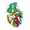
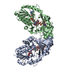
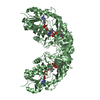
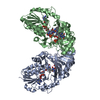
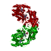
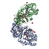
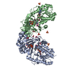
| Title | Structure of UDP-galactopyranose mutase bound to UDP-galactose (reduced) | ||||||
 Components Components | Probable UDP-galactopyranose mutase | ||||||
 Keywords Keywords | ISOMERASE / Flavoenzyme / protein-ligand complex / carbohydrate biosynthesis / FAD / Flavoprotein / Lipopolysaccharide biosynthesis | ||||||
| Function / homology |  Function and homology information Function and homology informationUDP-galactopyranose mutase / UDP-galactopyranose mutase activity / O antigen biosynthetic process / flavin adenine dinucleotide binding / cytosol Similarity search - Function | ||||||
| Biological species |  Klebsiella pneumoniae (bacteria) Klebsiella pneumoniae (bacteria) | ||||||
| Method |  X-RAY DIFFRACTION / SYNCHROTRON / MOLECULAR REPLACEMENT / Resolution: 2.51 Å X-RAY DIFFRACTION / SYNCHROTRON / MOLECULAR REPLACEMENT / Resolution: 2.51 Å | ||||||
 Authors Authors | Gruber, T.D. / Kiessling, L.L. / Forest, K.T. | ||||||
 Citation Citation | Journal: Biochemistry / Year: 2009 Title: X-ray crystallography reveals a reduced substrate complex of UDP-galactopyranose mutase poised for covalent catalysis by flavin . Authors: Gruber, T.D. / Westler, W.M. / Kiessling, L.L. / Forest, K.T. | ||||||
| History |
|
- Structure visualization
| Structure viewer | Molecule: MolmilJmol/JSmol |
|---|
- Downloads & links
-Download
| PDBx/mmCIF format | 3int.cif.gz | 175.4 KB | Display | PDBx/mmCIF format |
|---|---|---|---|---|
| PDB format | pdb3int.ent.gz | 137.7 KB | Display | PDB format |
| PDBx/mmJSON format | 3int.json.gz | Tree view | PDBx/mmJSON format | |
| Others |  Other downloads Other downloads |
-Validation report
| Arichive directory | https://data.pdbj.org/pub/pdb/validation_reports/in/3intftp://data.pdbj.org/pub/pdb/validation_reports/in/3int | HTTPS FTP |
|---|
-Related structure data
| Related structure data |  3inrC  3gf4S S: Starting model for refinement C: citing same article ( |
|---|---|
| Similar structure data |


-Links
 PDBj
PDBj


- Assembly
| Deposited unit | 
| ||||||||
|---|---|---|---|---|---|---|---|---|---|
| 1 |
| ||||||||
| 2 | 
| ||||||||
| 3 | 
| ||||||||
| Unit cell |
|
-Components
| #1: Protein | Mass: 45237.945 Da / Num. of mol.: 2 Source method: isolated from a genetically manipulated source Details: C-terminal Arg384 was altered to glycine during cloning, and six histidine residues were engineered on the C-terminus as a tag. Source: (gene. exp.) Klebsiella pneumoniae (bacteria) / Strain: 01 (ATCC 13882) / Gene: glf, rfbD / Plasmid: pGEM-Teasy / Production host: #2: Chemical |   Mass: 787.566 Da / Num. of mol.: 2 / Source method: obtained synthetically / Formula: C27H35N9O15P2 Mass: 787.566 Da / Num. of mol.: 2 / Source method: obtained synthetically / Formula: C27H35N9O15P2#3: Chemical | ChemComp-UDP / |   Type: RNA linking / Mass: 404.161 Da / Num. of mol.: 1 / Source method: obtained synthetically / Formula: C9H14N2O12P2 / Comment: UDP*YM Type: RNA linking / Mass: 404.161 Da / Num. of mol.: 1 / Source method: obtained synthetically / Formula: C9H14N2O12P2 / Comment: UDP*YM#4: Chemical | ChemComp-GDU / |   Mass: 566.302 Da / Num. of mol.: 1 Mass: 566.302 Da / Num. of mol.: 1Source method: isolated from a genetically manipulated source Formula: C15H24N2O17P2 #5: Water | ChemComp-HOH / |  Mass: 18.015 Da / Num. of mol.: 200 / Source method: isolated from a natural source / Formula: H2O Mass: 18.015 Da / Num. of mol.: 200 / Source method: isolated from a natural source / Formula: H2OSequence details | THESE ARE VERY CONSERVATIVE MUTATIONS FROM THE PUBLISHED SEQUENCE. THEY REFLECT SEQUENCE ...THESE ARE VERY CONSERVATI | |
|---|
-Experimental details
-Experiment
| Experiment | Method: X-RAY DIFFRACTION / Number of used crystals: 1 |
|---|
- Sample preparation
| Crystal | Density Matthews: 3.17 Å3/Da / Density % sol: 61.15 % |
|---|---|
| Crystal grow | Temperature: 298 K / Method: vapor diffusion, hanging drop / pH: 5.6 Details: Drops containing 1.5 microliters 5 mg/mL protein in 20 mM HEPES were combined with 1.5 microliters well solution (85 mM ammonium acetate, 42 mM tri-sodium citrate, 12.3% PEG 4000, 7.5% ...Details: Drops containing 1.5 microliters 5 mg/mL protein in 20 mM HEPES were combined with 1.5 microliters well solution (85 mM ammonium acetate, 42 mM tri-sodium citrate, 12.3% PEG 4000, 7.5% glycerol, 15 mM L-cysteine, 5 mM UDP-Glc) for 1-2 weeks. Crystals were then soaked in a solution of 53% Qiagen Cryos Suite Condition #87 with 15 mM L-cys, 30% methanol, 90 mM UDP-Galp (24hrs). Crystals were then soaked in a solution of 53% Qiagen Cryos Suite Condition #87 with 15 mM L-cys, 30% methanol, 90 mM UDP-Galp plus 100 mM sodium dithionite (3 min), pH 5.6, VAPOR DIFFUSION, HANGING DROP, temperature 298K |
-Data collection
| Diffraction | Mean temperature: 100 K |
|---|---|
| Diffraction source | Source: SYNCHROTRON / Site: APS  / Beamline: 21-ID-G / Wavelength: 0.97856 Å / Beamline: 21-ID-G / Wavelength: 0.97856 Å |
| Detector | Type: MARMOSAIC 300 mm CCD / Detector: CCD / Date: Nov 5, 2008 / Details: Beryllium lens |
| Radiation | Monochromator: C(111) diamond laue / Protocol: SINGLE WAVELENGTH / Monochromatic (M) / Laue (L): M / Scattering type: x-ray |
| Radiation wavelength | Wavelength: 0.97856 Å / Relative weight: 1 |
| Reflection | Resolution: 2.5→30 Å / Num. all: 38556 / Num. obs: 38510 / % possible obs: 100 % / Observed criterion σ(F): 0 / Observed criterion σ(I): 0 / Redundancy: 7.6 % / Rsym value: 0.107 / Net I/σ(I): 15 |
| Reflection shell | Resolution: 2.5→2.59 Å / Redundancy: 7.5 % / Mean I/σ(I) obs: 6.7 / Rsym value: 0.329 / % possible all: 100 |
- Processing
| Software |
| ||||||||||||||||||||||||||||||||||||||||||||||||||||||||||||||||||||||||||||||||||||||||||||||||||||||||||||||||||||||||||||||||||||||||||||||||||||||||||||||||||||||||||
|---|---|---|---|---|---|---|---|---|---|---|---|---|---|---|---|---|---|---|---|---|---|---|---|---|---|---|---|---|---|---|---|---|---|---|---|---|---|---|---|---|---|---|---|---|---|---|---|---|---|---|---|---|---|---|---|---|---|---|---|---|---|---|---|---|---|---|---|---|---|---|---|---|---|---|---|---|---|---|---|---|---|---|---|---|---|---|---|---|---|---|---|---|---|---|---|---|---|---|---|---|---|---|---|---|---|---|---|---|---|---|---|---|---|---|---|---|---|---|---|---|---|---|---|---|---|---|---|---|---|---|---|---|---|---|---|---|---|---|---|---|---|---|---|---|---|---|---|---|---|---|---|---|---|---|---|---|---|---|---|---|---|---|---|---|---|---|---|---|---|---|---|
| Refinement | Method to determine structure: MOLECULAR REPLACEMENT Starting model: 3GF4 Resolution: 2.51→30 Å / Cor.coef. Fo:Fc: 0.946 / Cor.coef. Fo:Fc free: 0.917 / SU B: 8.461 / SU ML: 0.19 / Cross valid method: THROUGHOUT / ESU R: 0.391 / ESU R Free: 0.267 / Stereochemistry target values: MAXIMUM LIKELIHOOD / Details: HYDROGENS HAVE BEEN ADDED IN THE RIDING POSITIONS
| ||||||||||||||||||||||||||||||||||||||||||||||||||||||||||||||||||||||||||||||||||||||||||||||||||||||||||||||||||||||||||||||||||||||||||||||||||||||||||||||||||||||||||
| Solvent computation | Ion probe radii: 0.8 Å / Shrinkage radii: 0.8 Å / VDW probe radii: 1.4 Å / Solvent model: MASK | ||||||||||||||||||||||||||||||||||||||||||||||||||||||||||||||||||||||||||||||||||||||||||||||||||||||||||||||||||||||||||||||||||||||||||||||||||||||||||||||||||||||||||
| Displacement parameters | Biso mean: 43.795 Å2
| ||||||||||||||||||||||||||||||||||||||||||||||||||||||||||||||||||||||||||||||||||||||||||||||||||||||||||||||||||||||||||||||||||||||||||||||||||||||||||||||||||||||||||
| Refine analyze |
| ||||||||||||||||||||||||||||||||||||||||||||||||||||||||||||||||||||||||||||||||||||||||||||||||||||||||||||||||||||||||||||||||||||||||||||||||||||||||||||||||||||||||||
| Refinement step | Cycle: LAST / Resolution: 2.51→30 Å
| ||||||||||||||||||||||||||||||||||||||||||||||||||||||||||||||||||||||||||||||||||||||||||||||||||||||||||||||||||||||||||||||||||||||||||||||||||||||||||||||||||||||||||
| Refine LS restraints |
| ||||||||||||||||||||||||||||||||||||||||||||||||||||||||||||||||||||||||||||||||||||||||||||||||||||||||||||||||||||||||||||||||||||||||||||||||||||||||||||||||||||||||||
| LS refinement shell | Resolution: 2.51→2.57 Å / Total num. of bins used: 20
|
