 Movie
Movie Controller

[English] 日本語
 Yorodumi
- PDB-2bi7: udp-galactopyranose mutase from Klebsiella pneumoniae oxidised FAD -
+ Open data

- Basic information
| Entry | Database: PDB / ID: 2bi7 | |||||||||
|---|---|---|---|---|---|---|---|---|---|---|
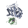
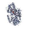
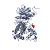
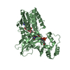
| Title | udp-galactopyranose mutase from Klebsiella pneumoniae oxidised FAD | |||||||||
 Components Components | UDP-GALACTOPYRANOSE MUTASE | |||||||||
 Keywords Keywords | ISOMERASE / FAD / FLAVOPROTEIN / LIPOPOLYSACCHARIDE BIOSYNTHESIS | |||||||||
| Function / homology |  Function and homology information Function and homology informationUDP-galactopyranose mutase / UDP-galactopyranose mutase activity / O antigen biosynthetic process / flavin adenine dinucleotide binding / cytosol Similarity search - Function | |||||||||
| Biological species |  KLEBSIELLA PNEUMONIAE (bacteria) KLEBSIELLA PNEUMONIAE (bacteria) | |||||||||
| Method |  X-RAY DIFFRACTION / MOLECULAR REPLACEMENT / Resolution: 2 Å X-RAY DIFFRACTION / MOLECULAR REPLACEMENT / Resolution: 2 Å | |||||||||
 Authors Authors | Beis, K. / Srikannathasan, V. / Naismith, J. | |||||||||
 Citation Citation | Journal: J.Mol.Biol. / Year: 2005 Title: Crystal Structures of Mycobacteria Tuberculosis and Klebsiella Pneumoniae Udp-Galactopyranose Mutase in the Oxidised State and Klebsiella Pneumoniae Udp-Galactopyranose Mutase in the (Active) Reduced State. Authors: Beis, K. / Srikannathasan, V. / Liu, H. / Fullerton, S.W.B. / Bamford, V.A. / Sanders, D.A.R. / Whitfield, C. / Mcneil, M.R. / Naismith, J.H. #1: Journal: Nat.Struct.Biol. / Year: 2001Title: Udp-Galactopyranose Mutase Has a Novel Structure and Mechanism Authors: Sanders, D.A. / Staines, A.G. / Mcmahon, S.A. / Mcneil, M.R. / Whitfield, C. / Naismith, J.H. | |||||||||
| History |
|
- Structure visualization
| Structure viewer | Molecule: MolmilJmol/JSmol |
|---|
- Downloads & links
-Download
| PDBx/mmCIF format | 2bi7.cif.gz | 95.5 KB | Display | PDBx/mmCIF format |
|---|---|---|---|---|
| PDB format | pdb2bi7.ent.gz | 71.4 KB | Display | PDB format |
| PDBx/mmJSON format | 2bi7.json.gz | Tree view | PDBx/mmJSON format | |
| Others |  Other downloads Other downloads |
-Validation report
| Arichive directory | https://data.pdbj.org/pub/pdb/validation_reports/bi/2bi7ftp://data.pdbj.org/pub/pdb/validation_reports/bi/2bi7 | HTTPS FTP |
|---|
-Related structure data
| Related structure data |  1v0jC  1wamC  2bi8C  1i8tS C: citing same article ( S: Starting model for refinement |
|---|---|
| Similar structure data |
-Links
 PDBj
PDBj- Assembly
| Deposited unit | 
| ||||||||
|---|---|---|---|---|---|---|---|---|---|
| 1 |
| ||||||||
| Unit cell |
|
-Components
| #1: Protein | Mass: 44512.180 Da / Num. of mol.: 1 Source method: isolated from a genetically manipulated source Source: (gene. exp.) KLEBSIELLA PNEUMONIAE (bacteria) / Production host: |
|---|---|
| #2: Chemical | ChemComp-FAD /   Mass: 785.550 Da / Num. of mol.: 1 / Source method: obtained synthetically / Formula: C27H33N9O15P2 / Comment: FAD*YM Mass: 785.550 Da / Num. of mol.: 1 / Source method: obtained synthetically / Formula: C27H33N9O15P2 / Comment: FAD*YM |
| #3: Water | ChemComp-HOH /  Mass: 18.015 Da / Num. of mol.: 140 / Source method: isolated from a natural source / Formula: H2O Mass: 18.015 Da / Num. of mol.: 140 / Source method: isolated from a natural source / Formula: H2O |
| Compound details | FUNCTION: INVOLVED IN THE CONVERSION |
-Experimental details
-Experiment
| Experiment | Method: X-RAY DIFFRACTION / Number of used crystals: 1 |
|---|
- Sample preparation
| Crystal | Density Matthews: 2.35 Å3/Da / Density % sol: 47 % |
|---|---|
| Crystal grow | pH: 6.5 Details: 0.1M BIS-TRIS PH6.5, 50MM CACL2, 30% PEG550, pH 6.50 |
-Data collection
| Diffraction | Mean temperature: 120 K |
|---|---|
| Diffraction source | Source: ROTATING ANODE / Type: RIGAKU MICROMAX-007 / Wavelength: 1.5418 |
| Detector | Type: RIGAKU RAXIS IV / Detector: IMAGE PLATE / Date: Jul 15, 2003 / Details: MIRRORS |
| Radiation | Protocol: SINGLE WAVELENGTH / Monochromatic (M) / Laue (L): M / Scattering type: x-ray |
| Radiation wavelength | Wavelength: 1.5418 Å / Relative weight: 1 |
| Reflection | Resolution: 2→74.54 Å / Num. obs: 2160 / % possible obs: 98.1 % / Redundancy: 16.6 % / Rmerge(I) obs: 0.05 / Net I/σ(I): 10.6 |
| Reflection shell | Resolution: 2→2.12 Å / Redundancy: 9.1 % / Rmerge(I) obs: 0.17 / Mean I/σ(I) obs: 4.3 / % possible all: 88.6 |
- Processing
| Software |
| ||||||||||||||||||||||||||||||||||||||||||||||||||||||||||||||||||||||||||||||||||||||||||||||||||||||||||||||||||||||||||||||||||||||||||||||||||||||||||||||||||||||||||||||||||||||
|---|---|---|---|---|---|---|---|---|---|---|---|---|---|---|---|---|---|---|---|---|---|---|---|---|---|---|---|---|---|---|---|---|---|---|---|---|---|---|---|---|---|---|---|---|---|---|---|---|---|---|---|---|---|---|---|---|---|---|---|---|---|---|---|---|---|---|---|---|---|---|---|---|---|---|---|---|---|---|---|---|---|---|---|---|---|---|---|---|---|---|---|---|---|---|---|---|---|---|---|---|---|---|---|---|---|---|---|---|---|---|---|---|---|---|---|---|---|---|---|---|---|---|---|---|---|---|---|---|---|---|---|---|---|---|---|---|---|---|---|---|---|---|---|---|---|---|---|---|---|---|---|---|---|---|---|---|---|---|---|---|---|---|---|---|---|---|---|---|---|---|---|---|---|---|---|---|---|---|---|---|---|---|---|
| Refinement | Method to determine structure: MOLECULAR REPLACEMENT Starting model: PDB ENTRY 1I8T Resolution: 2→74.54 Å / Cor.coef. Fo:Fc: 0.953 / Cor.coef. Fo:Fc free: 0.926 / SU ML: 0.127 / TLS residual ADP flag: LIKELY RESIDUAL / Cross valid method: THROUGHOUT / ESU R: 0.223 / ESU R Free: 0.194 / Stereochemistry target values: MAXIMUM LIKELIHOOD / Details: HYDROGENS HAVE BEEN ADDED IN THE RIDING POSITIONS.
| ||||||||||||||||||||||||||||||||||||||||||||||||||||||||||||||||||||||||||||||||||||||||||||||||||||||||||||||||||||||||||||||||||||||||||||||||||||||||||||||||||||||||||||||||||||||
| Solvent computation | Ion probe radii: 0.8 Å / Shrinkage radii: 0.8 Å / VDW probe radii: 1.2 Å / Solvent model: BABINET MODEL WITH MASK | ||||||||||||||||||||||||||||||||||||||||||||||||||||||||||||||||||||||||||||||||||||||||||||||||||||||||||||||||||||||||||||||||||||||||||||||||||||||||||||||||||||||||||||||||||||||
| Displacement parameters | Biso mean: 34.83 Å2
| ||||||||||||||||||||||||||||||||||||||||||||||||||||||||||||||||||||||||||||||||||||||||||||||||||||||||||||||||||||||||||||||||||||||||||||||||||||||||||||||||||||||||||||||||||||||
| Refinement step | Cycle: LAST / Resolution: 2→74.54 Å
| ||||||||||||||||||||||||||||||||||||||||||||||||||||||||||||||||||||||||||||||||||||||||||||||||||||||||||||||||||||||||||||||||||||||||||||||||||||||||||||||||||||||||||||||||||||||
| Refine LS restraints |
|