 Movie
Movie Controller


+ Open data

- Basic information
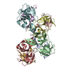
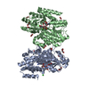
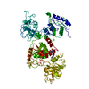
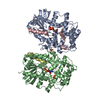
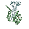
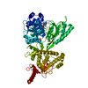
| Entry | Database: PDB / ID: 1i8t | ||||||
|---|---|---|---|---|---|---|---|
| Title | STRUCTURE OF UDP-GALACTOPYRANOSE MUTASE FROM E.COLI | ||||||
 Components Components | UDP-GALACTOPYRANOSE MUTASE | ||||||
 Keywords Keywords | ISOMERASE / Rossmann Fold / FAD / UDP-galactopyranose / mutase / contractase | ||||||
| Function / homology |  Function and homology information Function and homology informationUDP-galactopyranose mutase / UDP-galactopyranose mutase activity / lipopolysaccharide biosynthetic process / flavin adenine dinucleotide binding / protein homodimerization activity / cytosol Similarity search - Function | ||||||
| Biological species |  | ||||||
| Method |  X-RAY DIFFRACTION / SYNCHROTRON / MAD / Resolution: 2.4 Å X-RAY DIFFRACTION / SYNCHROTRON / MAD / Resolution: 2.4 Å | ||||||
 Authors Authors | Sanders, D.A.R. / Staines, A.G. / McMahon, S.A. / McNeil, M.R. / Whitfield, C. / Naismith, J.H. | ||||||
 Citation Citation | Journal: Nat.Struct.Biol. / Year: 2001 Title: UDP-galactopyranose mutase has a novel structure and mechanism. Authors: Sanders, D.A. / Staines, A.G. / McMahon, S.A. / McNeil, M.R. / Whitfield, C. / Naismith, J.H. #1: Journal: Acta Crystallogr.,Sect.D / Year: 2001Title: Molecular Placement of Experimental Electron Density: A Case Study on UDP-Galactopyranose Mutase Authors: Sanders, D.A.R. / McMahon, S.A. / Leonard, G.L. / Naimsith, J.H. | ||||||
| History |
|
- Structure visualization
| Structure viewer | Molecule: MolmilJmol/JSmol |
|---|
- Downloads & links
-Download
| PDBx/mmCIF format | 1i8t.cif.gz | 169.4 KB | Display | PDBx/mmCIF format |
|---|---|---|---|---|
| PDB format | pdb1i8t.ent.gz | 133.5 KB | Display | PDB format |
| PDBx/mmJSON format | 1i8t.json.gz | Tree view | PDBx/mmJSON format | |
| Others |  Other downloads Other downloads |
-Validation report
| Arichive directory | https://data.pdbj.org/pub/pdb/validation_reports/i8/1i8tftp://data.pdbj.org/pub/pdb/validation_reports/i8/1i8t | HTTPS FTP |
|---|
-Related structure data
| Similar structure data |
|---|


-Links
 PDBj
PDBj- Assembly
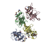
| Deposited unit | 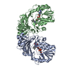
| ||||||||
|---|---|---|---|---|---|---|---|---|---|
| 1 |
| ||||||||
| Unit cell |
|
-Components
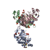
| #1: Protein | Mass: 43025.789 Da / Num. of mol.: 2 Source method: isolated from a genetically manipulated source Source: (gene. exp.) #2: Chemical |   Mass: 785.550 Da / Num. of mol.: 2 / Source method: obtained synthetically / Formula: C27H33N9O15P2 / Comment: FAD*YM Mass: 785.550 Da / Num. of mol.: 2 / Source method: obtained synthetically / Formula: C27H33N9O15P2 / Comment: FAD*YM#3: Water | ChemComp-HOH / |  Mass: 18.015 Da / Num. of mol.: 321 / Source method: isolated from a natural source / Formula: H2O Mass: 18.015 Da / Num. of mol.: 321 / Source method: isolated from a natural source / Formula: H2O |
|---|
-Experimental details
-Experiment
| Experiment | Method: X-RAY DIFFRACTION / Number of used crystals: 1 |
|---|
- Sample preparation
| Crystal | Density Matthews: 2.14 Å3/Da / Density % sol: 42.48 % | ||||||||||||||||||||||||||||||
|---|---|---|---|---|---|---|---|---|---|---|---|---|---|---|---|---|---|---|---|---|---|---|---|---|---|---|---|---|---|---|---|
| Crystal grow | Temperature: 298 K / Method: vapor diffusion, hanging drop / pH: 7.6 Details: PEG 4K, 0.01 mM l-cystein, Hepe, pH 7.6, VAPOR DIFFUSION, HANGING DROP, temperature 298K | ||||||||||||||||||||||||||||||
| Crystal grow | *PLUS pH: 7 / Method: vapor diffusionDetails: used macroseeding, Sanders, D.A.R., (2001) Acta Crystallogr., Sect.D, 57, 1415. | ||||||||||||||||||||||||||||||
| Components of the solutions | *PLUS
|
-Data collection
| Diffraction |
| |||||||||||||||
|---|---|---|---|---|---|---|---|---|---|---|---|---|---|---|---|---|
| Diffraction source | Source: SYNCHROTRON / Site: ESRF  / Beamline: ID14-2 / Wavelength: 0.934 Å / Beamline: ID14-2 / Wavelength: 0.934 Å | |||||||||||||||
| Detector | Type: MARRESEARCH / Detector: CCD / Date: Feb 28, 2000 | |||||||||||||||
| Radiation |
| |||||||||||||||
| Radiation wavelength | Wavelength: 0.934 Å / Relative weight: 1 | |||||||||||||||
| Reflection | Resolution: 2.4→40 Å / Num. all: 28865 / Num. obs: 28288 / % possible obs: 98 % / Observed criterion σ(F): 2 / Observed criterion σ(I): 2 / Redundancy: 3.9 % / Biso Wilson estimate: 37 Å2 / Rmerge(I) obs: 0.059 / Net I/σ(I): 10.9 | |||||||||||||||
| Reflection shell | Resolution: 2.4→2.52 Å / Redundancy: 3.7 % / Rmerge(I) obs: 0.224 / Mean I/σ(I) obs: 3.3 / % possible all: 93 | |||||||||||||||
| Reflection | *PLUS Lowest resolution: 40 Å / % possible obs: 98 % | |||||||||||||||
| Reflection shell | *PLUS % possible obs: 93 % |
- Processing
| Software |
| ||||||||||||||||||||
|---|---|---|---|---|---|---|---|---|---|---|---|---|---|---|---|---|---|---|---|---|---|
| Refinement | Method to determine structure: MAD / Resolution: 2.4→40 Å / Cross valid method: THROUGHOUT / σ(F): 2 / σ(I): 2 / Stereochemistry target values: Engh & Huber
| ||||||||||||||||||||
| Refinement step | Cycle: LAST / Resolution: 2.4→40 Å
| ||||||||||||||||||||
| LS refinement shell | Resolution: 2.4→2.52 Å | ||||||||||||||||||||
| Software | *PLUS Name: CNS / Version: 1 / Classification: refinement | ||||||||||||||||||||
| Refinement | *PLUS Highest resolution: 2.4 Å / Lowest resolution: 40 Å / σ(F): 2 / % reflection Rfree: 5 % / Rfactor obs: 0.185 | ||||||||||||||||||||
| Solvent computation | *PLUS | ||||||||||||||||||||
| Displacement parameters | *PLUS | ||||||||||||||||||||
| Refine LS restraints | *PLUS
| ||||||||||||||||||||
| LS refinement shell | *PLUS Highest resolution: 2.4 Å |
