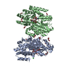
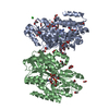
[English] 日本語
Yorodumi
- PDB-1aiv: APO OVOTRANSFERRIN -

+
Open data


ID or keywords:

Loading...

-
Basic information

Entry
Database: PDB / ID: 1aiv
TitleAPO OVOTRANSFERRIN
ComponentsOVOTRANSFERRIN
KeywordsIRON TRANSPORT PROTEIN
Function / homology
Function and homology information


extracellular sequestering of iron ion / organomineral extracellular matrix / antimicrobial humoral response / intracellular sequestering of iron ion / ferric iron binding / acute-phase response / recycling endosome / antibacterial humoral response / iron ion transport / response to lipopolysaccharide ...extracellular sequestering of iron ion / organomineral extracellular matrix / antimicrobial humoral response / intracellular sequestering of iron ion / ferric iron binding / acute-phase response / recycling endosome / antibacterial humoral response / iron ion transport / response to lipopolysaccharide / early endosome / response to xenobiotic stimulus / iron ion binding / extracellular space / plasma membrane
Similarity search - Function
Transferrin-like domain signature 2. / Transferrin family, iron binding site / Transferrin-like domain signature 1. / Transferrin-like domain signature 3. / Transferrin-like domain / Transferrin / Transferrin / Transferrin-like domain profile. / Transferrin / Periplasmic binding protein-like II ...Transferrin-like domain signature 2. / Transferrin family, iron binding site / Transferrin-like domain signature 1. / Transferrin-like domain signature 3. / Transferrin-like domain / Transferrin / Transferrin / Transferrin-like domain profile. / Transferrin / Periplasmic binding protein-like II / D-Maltodextrin-Binding Protein; domain 2 / 3-Layer(aba) Sandwich / Alpha Beta
Similarity search - Domain/homology
Biological speciesGallus gallus (chicken)
MethodX-RAY DIFFRACTION / MOLECULAR REPLACEMENT / Resolution: 3 Å
AuthorsKurokawa, H. / Dewan, J.C. / Mikami, B. / Sacchettini, J.C. / Hirose, M.
Citation
Journal: J.Biol.Chem. / Year: 1999
Title: Crystal structure of hen apo-ovotransferrin. Both lobes adopt an open conformation upon loss of iron
Authors: Kurokawa, H. / Dewan, J.C. / Mikami, B. / Sacchettini, J.C. / Hirose, M.
#1: Journal: J.Mol.Biol. / Year: 1995
Title: Crystal Structure of Diferric Hen Ovotransferrin at 2.4 A Resolution
Authors: Kurokawa, H. / Mikami, B. / Hirose, M.
History
DepositionApr 28, 1997Processing site: BNL
Revision 1.0Apr 29, 1998Provider: repository / Type: Initial release
Revision 1.1Mar 24, 2008Group: Version format compliance
Revision 1.2Jul 13, 2011Group: Non-polymer description / Version format compliance
Revision 2.0Jul 29, 2020Group: Atomic model / Data collection ...Atomic model / Data collection / Derived calculations / Structure summary
Category: atom_site / chem_comp ...atom_site / chem_comp / entity / pdbx_branch_scheme / pdbx_chem_comp_identifier / pdbx_entity_branch / pdbx_entity_branch_descriptor / pdbx_entity_branch_link / pdbx_entity_branch_list / pdbx_entity_nonpoly / pdbx_nonpoly_scheme / pdbx_struct_assembly_gen / struct_asym / struct_conn / struct_site / struct_site_gen
Item: _atom_site.auth_asym_id / _atom_site.auth_atom_id ..._atom_site.auth_asym_id / _atom_site.auth_atom_id / _atom_site.auth_seq_id / _atom_site.label_asym_id / _atom_site.label_atom_id / _atom_site.label_entity_id / _chem_comp.name / _chem_comp.type / _pdbx_struct_assembly_gen.asym_id_list / _struct_conn.pdbx_leaving_atom_flag / _struct_conn.pdbx_role / _struct_conn.ptnr1_auth_asym_id / _struct_conn.ptnr1_auth_comp_id / _struct_conn.ptnr1_auth_seq_id / _struct_conn.ptnr1_label_asym_id / _struct_conn.ptnr1_label_atom_id / _struct_conn.ptnr1_label_comp_id / _struct_conn.ptnr1_label_seq_id / _struct_conn.ptnr2_auth_asym_id / _struct_conn.ptnr2_auth_comp_id / _struct_conn.ptnr2_auth_seq_id / _struct_conn.ptnr2_label_asym_id / _struct_conn.ptnr2_label_atom_id / _struct_conn.ptnr2_label_comp_id / _struct_conn.ptnr2_label_seq_id
Description: Carbohydrate remediation / Provider: repository / Type: Remediation
Revision 2.1Aug 2, 2023Group: Database references / Refinement description / Structure summary
Category: chem_comp / database_2 / pdbx_initial_refinement_model
Item: _chem_comp.pdbx_synonyms / _database_2.pdbx_DOI / _database_2.pdbx_database_accession

-
Structure visualization

Structure viewerMolecule:
MolmilJmol/JSmol

Downloads & links

-
Assembly

Deposited unit
A: OVOTRANSFERRIN
hetero molecules


Theoretical massNumber of molelcules
Total (without water)76,3532
Polymers75,9291
Non-polymers4241
Water0
1


  • Idetical with deposited unit
  • defined by author
TypeNameSymmetry operationNumber
identity operation1_555x,y,z1
Unit cell
Length a, b, c (Å)92.260, 92.260, 178.190
Angle α, β, γ (deg.)90.00, 90.00, 90.00
Int Tables number96
Space group name H-MP43212

-
Components

#1: Protein OVOTRANSFERRIN / / CONALBUMIN


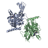
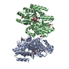
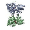
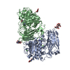
Mass: 75929.008 Da / Num. of mol.: 1 / Source method: isolated from a natural source / Details: APO FORM / Source: (natural) Gallus gallus (chicken) / References: UniProt: P02789
#2: Polysaccharide 2-acetamido-2-deoxy-alpha-D-glucopyranose-(1-4)-2-acetamido-2-deoxy-beta-D-glucopyranose


Type: oligosaccharide / Mass: 424.401 Da / Num. of mol.: 1
Source method: isolated from a genetically manipulated source
DescriptorTypeProgram
DGlcpNAca1-4DGlcpNAcb1-Glycam Condensed SequenceGMML 1.0
WURCS=2.0/2,2,1/[a2122h-1b_1-5_2*NCC/3=O][a2122h-1a_1-5_2*NCC/3=O]/1-2/a4-b1WURCSPDB2Glycan 1.1.0
[]{[(4+1)][b-D-GlcpNAc]{[(4+1)][a-D-GlcpNAc]{}}}LINUCSPDB-CARE
Sequence details
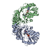
INTRASPECIFIC AND INTRA-INDIVIDUAL HETEROGENEITY OF OVOTRANSFERRIN HAS BEEN KNOWN FROM ...INTRASPECIFIC AND INTRA-INDIVIDUAL HETEROGENEITY OF OVOTRANSFERRIN HAS BEEN KNOWN FROM ELECTROPHORESIS STUDIES. THE DEPOSITORS' SEQUENCING STUDIES OF TRYPTIC FRAGMENTS OF OVOTRANSFERRIN USED FOR CRYSTALLOGRAPHIC STUDIES, SHOWED THAT RESIDUE 33 (29 IN ABOVE) IS SER OR ALA, THE RESIDUES 220 (216) AND 221 ARE GLN AND LEU, RESPECTIVELY, AND THE RESIDUE 135 IS ARG. THESE ARE CONSISTENT WITH THE SEQUENCE REPORTED BY JELTSCH, J. ET AL. (1987) NUCLEIC ACIDS RESEARCH 15, 7643-7645. THUS, THE DEPOSITORS DECIDED TO USE THE SEQUENCE. NO NEGATIVE OR POSITIVE FEATURE AROUND THE VARIABLE RESIDUES LISTED ABOVE CAN BE SEEN.

-
Experimental details

-
Experiment

ExperimentMethod: X-RAY DIFFRACTION / Number of used crystals: 1

-
Sample preparation

CrystalDensity Matthews: 2.5 Å3/Da / Density % sol: 50.72 %
Crystal growpH: 5.9 / Details: pH 5.9
Crystal grow
*PLUS
Temperature: 4 ℃ / pH: 6 / Method: vapor diffusion, hanging drop
Components of the solutions
*PLUS
IDConc.Common nameCrystal-IDSol-ID
142.5 mg/mlprotein1drop
20.02 Macetate1drop
32-3 %PEG60001drop
44-6 %PEG60001reservoir
50.02 Macetate1reservoir

-
Data collection

DiffractionMean temperature: 123 K
Diffraction sourceWavelength: 1.5418
DetectorType: SIEMENS / Detector: AREA DETECTOR / Date: Dec 20, 1996
RadiationMonochromator: COLLIMATOR / Monochromatic (M) / Laue (L): M / Scattering type: x-ray
Radiation wavelengthWavelength: 1.5418 Å / Relative weight: 1
ReflectionResolution: 3→20 Å / Num. obs: 12920 / % possible obs: 80.27 % / Observed criterion σ(I): 1 / Rsym value: 0.069 / Net I/σ(I): 6.5
Reflection
*PLUS
Num. measured all: 31370 / Rmerge(I) obs: 0.069

-
Processing

Software
NameVersionClassification
SAINTdata scaling
SAINTdata reduction
X-PLOR3.851model building
X-PLOR3.851refinement
X-PLOR3.851phasing
RefinementMethod to determine structure: MOLECULAR REPLACEMENT
Starting model: PDB ENTRY 1OVT
Resolution: 3→20 Å / Data cutoff high absF: 10000000 / Data cutoff low absF: 0.001 / Cross valid method: THROUGHOUT / σ(F): 2
RfactorNum. reflection% reflectionSelection details
Rfree0.265 --RANDOM
Rwork0.231 ---
obs0.231 11989 74.9 %-
Refinement stepCycle: LAST / Resolution: 3→20 Å
ProteinNucleic acidLigandSolventTotal
Num. atoms5312 0 28 0 5340
Refine LS restraints
Refine-IDTypeDev ideal
X-RAY DIFFRACTIONx_bond_d0.01
X-RAY DIFFRACTIONx_bond_d_na
X-RAY DIFFRACTIONx_bond_d_prot
X-RAY DIFFRACTIONx_angle_d
X-RAY DIFFRACTIONx_angle_d_na
X-RAY DIFFRACTIONx_angle_d_prot
X-RAY DIFFRACTIONx_angle_deg1.69
X-RAY DIFFRACTIONx_angle_deg_na
X-RAY DIFFRACTIONx_angle_deg_prot
X-RAY DIFFRACTIONx_dihedral_angle_d24.41
X-RAY DIFFRACTIONx_dihedral_angle_d_na
X-RAY DIFFRACTIONx_dihedral_angle_d_prot
X-RAY DIFFRACTIONx_improper_angle_d1.385
X-RAY DIFFRACTIONx_improper_angle_d_na
X-RAY DIFFRACTIONx_improper_angle_d_prot
X-RAY DIFFRACTIONx_mcbond_it
X-RAY DIFFRACTIONx_mcangle_it
X-RAY DIFFRACTIONx_scbond_it
X-RAY DIFFRACTIONx_scangle_it
Xplor file
Refine-IDSerial noParam fileTopol file
X-RAY DIFFRACTION1PARHCSDX.PROTOPH3.CHO
X-RAY DIFFRACTION2TOPHCSDX.PRO
Software
*PLUS
Name: X-PLOR / Version: 3.851 / Classification: refinement
Refine LS restraints
*PLUS
Refine-IDTypeDev ideal
X-RAY DIFFRACTIONx_dihedral_angle_d
X-RAY DIFFRACTIONx_dihedral_angle_deg24.413
X-RAY DIFFRACTIONx_improper_angle_d
X-RAY DIFFRACTIONx_improper_angle_deg1.385

+
About Yorodumi

-
News

-
Feb 9, 2022. New format data for meta-information of EMDB entries

New format data for meta-information of EMDB entries

  • Version 3 of the EMDB header file is now the official format.
  • The previous official version 1.9 will be removed from the archive.

Related info.:EMDB header

External links:wwPDB to switch to version 3 of the EMDB data model

-
Aug 12, 2020. Covid-19 info

Covid-19 info

URL: https://pdbj.org/emnavi/covid19.php

New page: Covid-19 featured information page in EM Navigator.

Related info.:Covid-19 info / Mar 5, 2020. Novel coronavirus structure data

+
Mar 5, 2020. Novel coronavirus structure data

Novel coronavirus structure data

Related info.:Yorodumi Speices / Aug 12, 2020. Covid-19 info

External links:COVID-19 featured content - PDBj / Molecule of the Month (242):Coronavirus Proteases

+
Jan 31, 2019. EMDB accession codes are about to change! (news from PDBe EMDB page)

EMDB accession codes are about to change! (news from PDBe EMDB page)

  • The allocation of 4 digits for EMDB accession codes will soon come to an end. Whilst these codes will remain in use, new EMDB accession codes will include an additional digit and will expand incrementally as the available range of codes is exhausted. The current 4-digit format prefixed with “EMD-” (i.e. EMD-XXXX) will advance to a 5-digit format (i.e. EMD-XXXXX), and so on. It is currently estimated that the 4-digit codes will be depleted around Spring 2019, at which point the 5-digit format will come into force.
  • The EM Navigator/Yorodumi systems omit the EMD- prefix.

Related info.:Q: What is EMD? / ID/Accession-code notation in Yorodumi/EM Navigator

External links:EMDB Accession Codes are Changing Soon! / Contact to PDBj

+
Jul 12, 2017. Major update of PDB

Major update of PDB

  • wwPDB released updated PDB data conforming to the new PDBx/mmCIF dictionary.
  • This is a major update changing the version number from 4 to 5, and with Remediation, in which all the entries are updated.
  • In this update, many items about electron microscopy experimental information are reorganized (e.g. em_software).
  • Now, EM Navigator and Yorodumi are based on the updated data.

External links:wwPDB Remediation / Enriched Model Files Conforming to OneDep Data Standards Now Available in the PDB FTP Archive

-
Yorodumi

Thousand views of thousand structures

  • Yorodumi is a browser for structure data from EMDB, PDB, SASBDB, etc.
  • This page is also the successor to EM Navigator detail page, and also detail information page/front-end page for Omokage search.
  • The word "yorodu" (or yorozu) is an old Japanese word meaning "ten thousand". "mi" (miru) is to see.

Related info.:EMDB / PDB / SASBDB / Comparison of 3 databanks / Yorodumi Search / Aug 31, 2016. New EM Navigator & Yorodumi / Yorodumi Papers / Jmol/JSmol / Function and homology information / Changes in new EM Navigator and Yorodumi

Read more