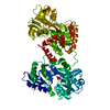
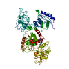
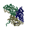
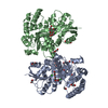
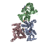
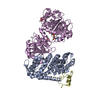
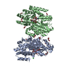
- PDB-1ryx: Crystal structure of hen serum transferrin in apo- form -
+
Open data
ID or keywords:
Loading...
-
Basic information
Entry
Database: PDB / ID: 1ryx
Title
Crystal structure of hen serum transferrin in apo- form
Components
Ovotransferrin
Keywords
METAL TRANSPORT / Hen serum transferrin / Apo- form / Domain Orientation
Function / homology
Function and homology information
organomineral extracellular matrix / iron ion transmembrane transport / antimicrobial humoral response / ferric iron binding / acute-phase response / iron ion transport / recycling endosome / antibacterial humoral response / response to lipopolysaccharide / intracellular iron ion homeostasis ...organomineral extracellular matrix / iron ion transmembrane transport / antimicrobial humoral response / ferric iron binding / acute-phase response / iron ion transport / recycling endosome / antibacterial humoral response / response to lipopolysaccharide / intracellular iron ion homeostasis / early endosome / iron ion binding / response to xenobiotic stimulus / : / plasma membrane Similarity search - Function
Resolution: 3.5→19.69 Å / Rfactor Rfree error: 0.016 / Data cutoff high absF: 4475883.76 / Data cutoff low absF: 0 / Isotropic thermal model: OVERALL / Cross valid method: THROUGHOUT / σ(F): 0 / Stereochemistry target values: Engh & Huber Details: THE RESIDUES WHOSE SIDE CHAINS COULD NOT BE LOCATED IN THE ELECTRON DENSITY MAP WERE KEPT AS ALANINE, HOWEVER, THE RESIDUE NAMES REMAIN UNALTERED IN THIS PDB FILE.
Rfactor
Num. reflection
% reflection
Selection details
Rfree
0.341
444
5.2 %
RANDOM
Rwork
0.283
-
-
-
obs
0.283
8604
88.3 %
-
Displacement parameters
Biso mean: 21.6 Å2
Baniso -1
Baniso -2
Baniso -3
1-
2.89 Å2
0 Å2
0 Å2
2-
-
2.89 Å2
0 Å2
3-
-
-
-5.78 Å2
Refine analyze
Free
Obs
Luzzati coordinate error
0.75 Å
0.58 Å
Luzzati d res low
-
20 Å
Luzzati sigma a
0.66 Å
0.42 Å
Refinement step
Cycle: LAST / Resolution: 3.5→19.69 Å
Protein
Nucleic acid
Ligand
Solvent
Total
Num. atoms
4657
0
0
0
4657
Refine LS restraints
Refine-ID
Type
Dev ideal
X-RAY DIFFRACTION
c_bond_d
0.011
X-RAY DIFFRACTION
c_angle_deg
1.9
X-RAY DIFFRACTION
c_dihedral_angle_d
25.2
X-RAY DIFFRACTION
c_improper_angle_d
1.29
LS refinement shell
Resolution: 3.5→3.72 Å / Rfactor Rfree error: 0.051 / Total num. of bins used: 6
In the structure databanks used in Yorodumi, some data are registered as the other names, "COVID-19 virus" and "2019-nCoV". Here are the details of the virus and the list of structure data.
Jan 31, 2019. EMDB accession codes are about to change! (news from PDBe EMDB page)
EMDB accession codes are about to change! (news from PDBe EMDB page)
The allocation of 4 digits for EMDB accession codes will soon come to an end. Whilst these codes will remain in use, new EMDB accession codes will include an additional digit and will expand incrementally as the available range of codes is exhausted. The current 4-digit format prefixed with “EMD-” (i.e. EMD-XXXX) will advance to a 5-digit format (i.e. EMD-XXXXX), and so on. It is currently estimated that the 4-digit codes will be depleted around Spring 2019, at which point the 5-digit format will come into force.
The EM Navigator/Yorodumi systems omit the EMD- prefix.
Related info.:Q: What is EMD? / ID/Accession-code notation in Yorodumi/EM Navigator
Yorodumi is a browser for structure data from EMDB, PDB, SASBDB, etc.
This page is also the successor to EM Navigator detail page, and also detail information page/front-end page for Omokage search.
The word "yorodu" (or yorozu) is an old Japanese word meaning "ten thousand". "mi" (miru) is to see.
Related info.:EMDB / PDB / SASBDB / Comparison of 3 databanks / Yorodumi Search / Aug 31, 2016. New EM Navigator & Yorodumi / Yorodumi Papers / Jmol/JSmol / Function and homology information / Changes in new EM Navigator and Yorodumi
 Movie
Movie Controller


 Open data
 Basic information
 Components
 Keywords
 Function and homology information
 X-RAY DIFFRACTION /
  Authors
 Citation
 Structure visualization
 Downloads & links
 Other downloads


 PDBj
PDBj


 Assembly
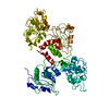
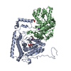
 Sample preparation
 Processing