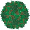
Movie
Movie Controller


+ Open data

- Basic information
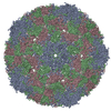
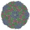
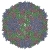
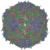
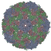
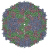
| Entry | Database: PDB / ID: 3j48 | ||||||
|---|---|---|---|---|---|---|---|
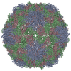
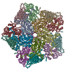
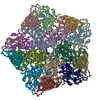
| Title | Cryo-EM structure of Poliovirus 135S particles | ||||||
 Components Components |
| ||||||
 Keywords Keywords | VIRUS / cell entry / single particle analysis | ||||||
| Function / homology |  Function and homology information Function and homology informationsymbiont-mediated suppression of host translation initiation / symbiont-mediated suppression of host cytoplasmic pattern recognition receptor signaling pathway via inhibition of RIG-I activity / symbiont-mediated suppression of host cytoplasmic pattern recognition receptor signaling pathway via inhibition of MDA-5 activity / symbiont-mediated suppression of host cytoplasmic pattern recognition receptor signaling pathway via inhibition of MAVS activity / picornain 2A / symbiont-mediated suppression of host mRNA export from nucleus / symbiont genome entry into host cell via pore formation in plasma membrane / picornain 3C / T=pseudo3 icosahedral viral capsid / host cell cytoplasmic vesicle membrane ...symbiont-mediated suppression of host translation initiation / symbiont-mediated suppression of host cytoplasmic pattern recognition receptor signaling pathway via inhibition of RIG-I activity / symbiont-mediated suppression of host cytoplasmic pattern recognition receptor signaling pathway via inhibition of MDA-5 activity / symbiont-mediated suppression of host cytoplasmic pattern recognition receptor signaling pathway via inhibition of MAVS activity / picornain 2A / symbiont-mediated suppression of host mRNA export from nucleus / symbiont genome entry into host cell via pore formation in plasma membrane / picornain 3C / T=pseudo3 icosahedral viral capsid / host cell cytoplasmic vesicle membrane / ribonucleoside triphosphate phosphatase activity / nucleoside-triphosphate phosphatase / channel activity / monoatomic ion transmembrane transport / RNA helicase activity / endocytosis involved in viral entry into host cell / symbiont-mediated activation of host autophagy / RNA-directed RNA polymerase / cysteine-type endopeptidase activity / viral RNA genome replication / RNA-directed RNA polymerase activity / virion attachment to host cell / DNA-templated transcription / host cell nucleus / structural molecule activity / proteolysis / RNA binding / zinc ion binding / ATP binding Similarity search - Function | ||||||
| Biological species |  Human poliovirus 1 Human poliovirus 1 | ||||||
| Method | ELECTRON MICROSCOPY / single particle reconstruction / cryo EM / Resolution: 5.5 Å | ||||||
 Authors Authors | Butan, C. / Fiman, D.J. / Hogle, J.M. | ||||||
 Citation Citation | Journal: J Virol / Year: 2014 Title: Cryo-electron microscopy reconstruction shows poliovirus 135S particles poised for membrane interaction and RNA release. Authors: Carmen Butan / David J Filman / James M Hogle /  Abstract: During infection, binding of mature poliovirus to cell surface receptors induces an irreversible expansion of the capsid, to form an infectious cell-entry intermediate particle that sediments at 135S. ...During infection, binding of mature poliovirus to cell surface receptors induces an irreversible expansion of the capsid, to form an infectious cell-entry intermediate particle that sediments at 135S. In these expanded virions, the major capsid proteins (VP1 to VP3) adopt an altered icosahedral arrangement to open holes in the capsid at 2-fold and quasi-3-fold axes, and internal polypeptides VP4 and the N terminus of VP1, which can bind membranes, become externalized. Cryo-electron microscopy images for 117,330 particles were collected using Leginon and reconstructed using FREALIGN. Improved rigid-body positioning of major capsid proteins established reliably which polypeptide segments become disordered or rearranged. The virus-to-135S transition includes expansion of 4%, rearrangements of the GH loops of VP3 and VP1, and disordering of C-terminal extensions of VP1 and VP2. The N terminus of VP1 rearranges to become externalized near its quasi-3-fold exit, binds to rearranged GH loops of VP3 and VP1, and attaches to the top surface of VP2. These details improve our understanding of subsequent stages of infection, including endocytosis and RNA transfer into the cytoplasm. | ||||||
| History |
|
- Structure visualization
| Movie |
Movie viewer |
|---|---|
| Structure viewer | Molecule: MolmilJmol/JSmol |
- Downloads & links
-Download
| PDBx/mmCIF format | 3j48.cif.gz | 38.4 KB | Display | PDBx/mmCIF format |
|---|---|---|---|---|
| PDB format | pdb3j48.ent.gz | 18.6 KB | Display | PDB format |
| PDBx/mmJSON format | 3j48.json.gz | Tree view | PDBx/mmJSON format | |
| Others |  Other downloads Other downloads |
-Validation report
| Arichive directory | https://data.pdbj.org/pub/pdb/validation_reports/j4/3j48ftp://data.pdbj.org/pub/pdb/validation_reports/j4/3j48 | HTTPS FTP |
|---|
-Related structure data
| Related structure data | 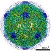 5710MC M: map data used to model this data C: citing same article ( |
|---|---|
| Similar structure data |
-Links
 PDBj
PDBj

- Assembly
| Deposited unit | 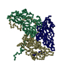
|
|---|---|
| 1 | x 60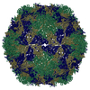
|
| 2 |
|
| 3 | x 5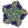
|
| 4 | x 6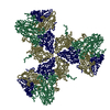
|
| 5 | 
|
| Symmetry | Point symmetry: (Schoenflies symbol: I (icosahedral)) |
-Components
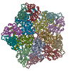
| #1: Protein | Mass: 33488.613 Da / Num. of mol.: 1 / Fragment: UNP residues 580-881 Source method: isolated from a genetically manipulated source Source: (gene. exp.) Human poliovirus 1 / Strain: Mahoney / Cell line (production host): HeLa / Production host:  Homo sapiens (human) / References: UniProt: P03300 Homo sapiens (human) / References: UniProt: P03300 |
|---|---|
| #2: Protein | Mass: 30075.783 Da / Num. of mol.: 1 / Fragment: UNP residues 70-341 Source method: isolated from a genetically manipulated source Source: (gene. exp.) Human poliovirus 1 / Strain: Mahoney / Cell line (production host): HeLa / Production host: Homo sapiens (human) / References: UniProt: P03300 |
| #3: Protein | Mass: 26547.482 Da / Num. of mol.: 1 / Fragment: UNP residues 342-579 Source method: isolated from a genetically manipulated source Source: (gene. exp.) Human poliovirus 1 / Strain: Mahoney / Cell line (production host): HeLa / Production host: Homo sapiens (human) / References: UniProt: P03300 |
-Experimental details
-Experiment
| Experiment | Method: ELECTRON MICROSCOPY |
|---|---|
| EM experiment | Aggregation state: PARTICLE / 3D reconstruction method: single particle reconstruction |
- Sample preparation
| Component | Name: Poliovirus 1 Mahoney 135S particle / Type: VIRUS Details: icosahedrally ordered capsid: 60 copies of VP1, VP2, VP3 | ||||||||||||
|---|---|---|---|---|---|---|---|---|---|---|---|---|---|
| Molecular weight |
| ||||||||||||
| Details of virus | Empty: NO / Enveloped: NO / Host category: VERTEBRATES / Isolate: STRAIN / Type: VIRION | ||||||||||||
| Natural host | Organism: Homo sapiens | ||||||||||||
| Buffer solution | Name: 20 mM HEPES, 2 mM CaCl2 / pH: 7.4 / Details: 20 mM HEPES, 2 mM CaCl2 | ||||||||||||
| Specimen | Conc.: 0.3 mg/ml / Embedding applied: NO / Shadowing applied: NO / Staining applied: NO / Vitrification applied: YES | ||||||||||||
| Specimen support | Details: glow-discharged holey carbon-grids (200 mesh C-flat grids) | ||||||||||||
| Vitrification | Instrument: HOMEMADE PLUNGER / Cryogen name: ETHANE / Temp: 90 K Details: Blotted manually in ambient atmosphere before plunging into ethane cooled by liquid nitrogen. Method: Blotted manually before plunge freezing into liquid ethane |
- Electron microscopy imaging
| Experimental equipment |  Model: Tecnai F20 / Image courtesy: FEI Company |
|---|---|
| Microscopy | Model: FEI TECNAI F20 / Date: Feb 28, 2011 |
| Electron gun | Electron source:  FIELD EMISSION GUN / Accelerating voltage: 200 kV / Illumination mode: FLOOD BEAM FIELD EMISSION GUN / Accelerating voltage: 200 kV / Illumination mode: FLOOD BEAM |
| Electron lens | Mode: BRIGHT FIELD / Nominal magnification: 62000 X / Nominal defocus max: 2000 nm / Nominal defocus min: 980 nm / Cs: 2 mm / Astigmatism: Objective lens astigmatism was corrected |
| Specimen holder | Specimen holder model: GATAN LIQUID NITROGEN Specimen holder type: Side entry liquid nitrogen-cooled cryo specimen holder Temperature: 90 K / Temperature (max): 93 K / Temperature (min): 90 K / Tilt angle max: 0 ° / Tilt angle min: 0 ° |
| Image recording | Electron dose: 15 e/Å2 / Film or detector model: TVIPS TEMCAM-F415 (4k x 4k) |
| Image scans | Num. digital images: 1020 |
| Radiation | Protocol: SINGLE WAVELENGTH / Monochromatic (M) / Laue (L): M / Scattering type: x-ray |
| Radiation wavelength | Relative weight: 1 |
- Processing
| EM software |
| ||||||||||||
|---|---|---|---|---|---|---|---|---|---|---|---|---|---|
| CTF correction | Details: Each micrograph | ||||||||||||
| Symmetry | Point symmetry: I (icosahedral) | ||||||||||||
| 3D reconstruction | Method: FREALIGN randomized search / Resolution: 5.5 Å / Resolution method: FSC 0.143 CUT-OFF / Num. of particles: 117330 / Nominal pixel size: 1.37 Å / Actual pixel size: 1.37 Å Details: Single particle details: The particles were selected using an automatic selection program (Single particle--Applied symmetry: I) Symmetry type: POINT | ||||||||||||
| Atomic model building | Protocol: RIGID BODY FIT / Space: RECIPROCAL Target criteria: mean amplitude-weighted cosine of the phase difference Details: METHOD--rigid body REFINEMENT PROTOCOL--rigid body DETAILS--Most of the model was docked, with specific areas of discrepancy fitted. The fitting was rigid body with flexible fitting or ...Details: METHOD--rigid body REFINEMENT PROTOCOL--rigid body DETAILS--Most of the model was docked, with specific areas of discrepancy fitted. The fitting was rigid body with flexible fitting or deletion of selected polypeptide segments. Rigid bodies for VP1, VP2, VP3, and the VP3 beta tube were defined to include beta barrels and non-covalently attached polypeptides. Each rigid body was repeatedly fitted manually and then refined. Disordered polypeptide segments were removed. Several rearranged segments were included as approximate backbone traces and refined. | ||||||||||||
| Atomic model building | 3D fitting-ID: 1 / Accession code: 1POV / Initial refinement model-ID: 1 / PDB-ID: 1POV / Source name: PDB / Type: experimental model
| ||||||||||||
| Refinement step | Cycle: LAST
|