 Movie
Movie Controller


+ Open data

- Basic information
| Entry | Database: PDB / ID: 4rr3 | ||||||
|---|---|---|---|---|---|---|---|
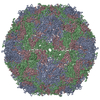
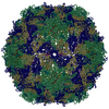
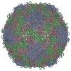
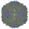
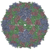
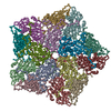
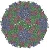
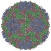
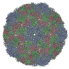
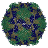
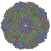
| Title | Crystal structure of a recombinant EV71 virus particle | ||||||
 Components Components |
| ||||||
 Keywords Keywords | VIRUS / beta barrel / eight-stranded beta barrel / replicate in the cytoplasm | ||||||
| Function / homology |  Function and homology information Function and homology informationsymbiont-mediated suppression of host cytoplasmic pattern recognition receptor signaling pathway via inhibition of MDA-5 activity / picornain 2A / symbiont-mediated suppression of host mRNA export from nucleus / symbiont genome entry into host cell via pore formation in plasma membrane / picornain 3C / T=pseudo3 icosahedral viral capsid / host cell cytoplasmic vesicle membrane / ribonucleoside triphosphate phosphatase activity / nucleoside-triphosphate phosphatase / channel activity ...symbiont-mediated suppression of host cytoplasmic pattern recognition receptor signaling pathway via inhibition of MDA-5 activity / picornain 2A / symbiont-mediated suppression of host mRNA export from nucleus / symbiont genome entry into host cell via pore formation in plasma membrane / picornain 3C / T=pseudo3 icosahedral viral capsid / host cell cytoplasmic vesicle membrane / ribonucleoside triphosphate phosphatase activity / nucleoside-triphosphate phosphatase / channel activity / monoatomic ion transmembrane transport / DNA replication / RNA helicase activity / endocytosis involved in viral entry into host cell / symbiont-mediated activation of host autophagy / RNA-directed RNA polymerase / cysteine-type endopeptidase activity / viral RNA genome replication / RNA-directed RNA polymerase activity / virion attachment to host cell / host cell nucleus / DNA-templated transcription / structural molecule activity / proteolysis / RNA binding / zinc ion binding / ATP binding Similarity search - Function | ||||||
| Biological species |   Enterovirus A71 Enterovirus A71 | ||||||
| Method |  X-RAY DIFFRACTION / SYNCHROTRON / MOLECULAR REPLACEMENT / Resolution: 3.103 Å X-RAY DIFFRACTION / SYNCHROTRON / MOLECULAR REPLACEMENT / Resolution: 3.103 Å | ||||||
 Authors Authors | Chen, R. / Lyu, K. | ||||||
 Citation Citation | Journal: J.Biol.Chem. / Year: 2015 Title: Crystal structures of enterovirus 71 (EV71) recombinant virus particles provide insights into vaccine design. Authors: Lyu, K. / Wang, G.C. / He, Y.L. / Han, J.F. / Ye, Q. / Qin, C.F. / Chen, R. | ||||||
| History |
|
- Structure visualization
| Structure viewer | Molecule: MolmilJmol/JSmol |
|---|
- Downloads & links
-Download
| PDBx/mmCIF format | 4rr3.cif.gz | 675.2 KB | Display | PDBx/mmCIF format |
|---|---|---|---|---|
| PDB format | pdb4rr3.ent.gz | 555 KB | Display | PDB format |
| PDBx/mmJSON format | 4rr3.json.gz | Tree view | PDBx/mmJSON format | |
| Others |  Other downloads Other downloads |
-Validation report
| Arichive directory | https://data.pdbj.org/pub/pdb/validation_reports/rr/4rr3ftp://data.pdbj.org/pub/pdb/validation_reports/rr/4rr3 | HTTPS FTP |
|---|
-Related structure data


-Links
 PDBj
PDBj

- Assembly
| Deposited unit | 
| ||||||||
|---|---|---|---|---|---|---|---|---|---|
| 1 | x 12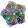
| ||||||||
| Unit cell |
| ||||||||
| Details | The biological assembly is a 60mer generated from the pentamer in the asymmetric unit by the following 12 operations: (x,y,z),(-x,-y,z),(-x,y,-z),(x,-y,-z),(y,z,x),(-y,-z,x),(-y,z,-x),(y,-z,-x),(z,x,y),(-z,-x,y),(-z,x,-y) and (z,-x,-y). The pentamer in the asymmetric unit is generated from the deposited PDB by the following transformations(r11,r12,r13,r21,r22,r23,r31,r32,r33,tx,tx,tz):(1,0,0,0,1,0,0,0,1,0,0,0), (0.3038, -0.8069, 0.5066, 0.804, 0.5025, 0.3181, -0.5112, 0.3106, 0.8014, -0.9474, -1.348, 0.9465), (-0.8079, 0.4819, -0.3392, -0.5134, -0.2927, 0.8067, 0.2895, 0.8259, 0.4839, 5.429, -0.9913, 0.9334), (-0.8132, -0.5063, 0.287, 0.4827, -0.3113, 0.8186, -0.3251, 0.8042, 0.4975, 3.379, -1.134, 0.6254) and (0.3056, 0.8031, -0.5116, -0.8063, 0.504, 0.3097, 0.5065, 0.3179, 0.8015, 2.154, -0.4491, 0.1634). |
-Components
| #1: Protein | Mass: 33499.781 Da / Num. of mol.: 5 / Mutation: K550Q Source method: isolated from a genetically manipulated source Source: (gene. exp.) Enterovirus A71 / Cell line (production host): RD cells / Production host:  Homo sapiens (human) / References: UniProt: F6KTB0 Homo sapiens (human) / References: UniProt: F6KTB0#2: Protein | Mass: 26440.148 Da / Num. of mol.: 5 Source method: isolated from a genetically manipulated source Source: (gene. exp.) Enterovirus A71 / Cell line (production host): RD cells / Production host: Homo sapiens (human) / References: UniProt: F6KTB0#3: Protein | Mass: 35209.219 Da / Num. of mol.: 5 Source method: isolated from a genetically manipulated source Source: (gene. exp.) Enterovirus A71 / Cell line (production host): RD cells / Production host: Homo sapiens (human) / References: UniProt: F6KTB0 |
|---|
-Experimental details
-Experiment
| Experiment | Method: X-RAY DIFFRACTION / Number of used crystals: 1 |
|---|
- Sample preparation
| Crystal | Density Matthews: 3.77 Å3/Da / Density % sol: 67.41 % |
|---|---|
| Crystal grow | Temperature: 290 K / Method: vapor diffusion, hanging drop / pH: 6.5 Details: 0.1M Imidazole containing 1M sodium acetate, pH 6.5, VAPOR DIFFUSION, HANGING DROP, temperature 290K |
-Data collection
| Diffraction | Mean temperature: 90 K |
|---|---|
| Diffraction source | Source: SYNCHROTRON / Site: SSRF  / Beamline: BL17U / Wavelength: 0.97803 Å / Beamline: BL17U / Wavelength: 0.97803 Å |
| Detector | Type: ADSC QUANTUM 315 / Detector: CCD / Date: Jun 6, 2012 |
| Radiation | Monochromator: SI 111 CHANNEL / Protocol: SINGLE WAVELENGTH / Monochromatic (M) / Laue (L): M / Scattering type: x-ray |
| Radiation wavelength | Wavelength: 0.97803 Å / Relative weight: 1 |
| Reflection | Resolution: 3.1→50 Å / Num. all: 131863 / % possible obs: 90.72 % / Observed criterion σ(F): 0 / Observed criterion σ(I): 0 |
| Reflection shell | Resolution: 3.1→3.15 Å / Redundancy: 6.7 % / Rmerge(I) obs: 0.967 / Mean I/σ(I) obs: 2.5 / Num. unique all: 6498 / % possible all: 100 |
- Processing
| Software |
| ||||||||||||||||||||
|---|---|---|---|---|---|---|---|---|---|---|---|---|---|---|---|---|---|---|---|---|---|
| Refinement | Method to determine structure: MOLECULAR REPLACEMENT / Resolution: 3.103→46.438 Å / σ(F): 1.33 / Stereochemistry target values: Engh & Huber
| ||||||||||||||||||||
| Refinement step | Cycle: LAST / Resolution: 3.103→46.438 Å
| ||||||||||||||||||||
| Refine LS restraints |
| ||||||||||||||||||||
| LS refinement shell | Resolution: 3.1034→3.181 Å
|
