 Movie
Movie Controller

[English] 日本語
 Yorodumi
- PDB-4n53: Human enterovirus 71 uncoating intermediate captured at atomic re... -
+ Open data

- Basic information
| Entry | Database: PDB / ID: 4n53 | ||||||
|---|---|---|---|---|---|---|---|
| Title | Human enterovirus 71 uncoating intermediate captured at atomic resolution | ||||||
 Components Components |
| ||||||
 Keywords Keywords | VIRUS / hand-foot-and-mouth disease / human enterovirus 71 / virion / pocket factor / picornavirus / icosahedral virus | ||||||
| Function / homology |  Function and homology information Function and homology information: / symbiont-mediated suppression of host cytoplasmic pattern recognition receptor signaling pathway via inhibition of MDA-5 activity / picornain 2A / symbiont-mediated suppression of host mRNA export from nucleus / symbiont genome entry into host cell via pore formation in plasma membrane / picornain 3C / T=pseudo3 icosahedral viral capsid / host cell cytoplasmic vesicle membrane / virion component / viral capsid ...: / symbiont-mediated suppression of host cytoplasmic pattern recognition receptor signaling pathway via inhibition of MDA-5 activity / picornain 2A / symbiont-mediated suppression of host mRNA export from nucleus / symbiont genome entry into host cell via pore formation in plasma membrane / picornain 3C / T=pseudo3 icosahedral viral capsid / host cell cytoplasmic vesicle membrane / virion component / viral capsid / host cell / nucleoside-triphosphate phosphatase / channel activity / monoatomic ion transmembrane transport / DNA replication / RNA helicase activity / endocytosis involved in viral entry into host cell / symbiont-mediated suppression of host gene expression / symbiont-mediated activation of host autophagy / RNA-directed RNA polymerase / cysteine-type endopeptidase activity / viral RNA genome replication / RNA-directed RNA polymerase activity / symbiont entry into host cell / virion attachment to host cell / DNA-templated transcription / host cell nucleus / structural molecule activity / ATP hydrolysis activity / proteolysis / RNA binding / ATP binding / membrane / metal ion binding Similarity search - Function | ||||||
| Biological species |   Enterovirus A71 Enterovirus A71 | ||||||
| Method |  X-RAY DIFFRACTION / SYNCHROTRON / MOLECULAR REPLACEMENT / Resolution: 3.3063 Å X-RAY DIFFRACTION / SYNCHROTRON / MOLECULAR REPLACEMENT / Resolution: 3.3063 Å | ||||||
 Authors Authors | Chen, R. / Lyu, K. | ||||||
 Citation Citation | Journal: J.Virol. / Year: 2014 Title: Human enterovirus 71 uncoating captured at atomic resolution. Authors: Lyu, K. / Ding, J. / Han, J.F. / Zhang, Y. / Wu, X.Y. / He, Y.L. / Qin, C.F. / Chen, R. | ||||||
| History |
|
- Structure visualization
| Structure viewer | Molecule: MolmilJmol/JSmol |
|---|
- Downloads & links
-Download
| PDBx/mmCIF format | 4n53.cif.gz | 176.6 KB | Display | PDBx/mmCIF format |
|---|---|---|---|---|
| PDB format | pdb4n53.ent.gz | 139.6 KB | Display | PDB format |
| PDBx/mmJSON format | 4n53.json.gz | Tree view | PDBx/mmJSON format | |
| Others |  Other downloads Other downloads |
-Validation report
| Arichive directory | https://data.pdbj.org/pub/pdb/validation_reports/n5/4n53ftp://data.pdbj.org/pub/pdb/validation_reports/n5/4n53 | HTTPS FTP |
|---|
-Related structure data
| Related structure data |  4n43C 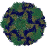 1d4mS C: citing same article ( S: Starting model for refinement |
|---|---|
| Similar structure data |


-Links
 PDBj
PDBj

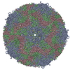
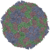
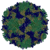
- Assembly
| Deposited unit | 
| ||||||||||||||||||||||||||||||||||||||||||||||||||||||||||||||||||||||||||||||||||||
|---|---|---|---|---|---|---|---|---|---|---|---|---|---|---|---|---|---|---|---|---|---|---|---|---|---|---|---|---|---|---|---|---|---|---|---|---|---|---|---|---|---|---|---|---|---|---|---|---|---|---|---|---|---|---|---|---|---|---|---|---|---|---|---|---|---|---|---|---|---|---|---|---|---|---|---|---|---|---|---|---|---|---|---|---|---|
| 1 | x 60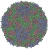
| ||||||||||||||||||||||||||||||||||||||||||||||||||||||||||||||||||||||||||||||||||||
| 2 |
| ||||||||||||||||||||||||||||||||||||||||||||||||||||||||||||||||||||||||||||||||||||
| 3 | x 5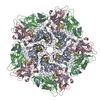
| ||||||||||||||||||||||||||||||||||||||||||||||||||||||||||||||||||||||||||||||||||||
| 4 | x 6
| ||||||||||||||||||||||||||||||||||||||||||||||||||||||||||||||||||||||||||||||||||||
| 5 | 
| ||||||||||||||||||||||||||||||||||||||||||||||||||||||||||||||||||||||||||||||||||||
| 6 | x 20
| ||||||||||||||||||||||||||||||||||||||||||||||||||||||||||||||||||||||||||||||||||||
| Unit cell |
| ||||||||||||||||||||||||||||||||||||||||||||||||||||||||||||||||||||||||||||||||||||
| Symmetry | Point symmetry: (Schoenflies symbol: I (icosahedral)) | ||||||||||||||||||||||||||||||||||||||||||||||||||||||||||||||||||||||||||||||||||||
| Noncrystallographic symmetry (NCS) | NCS oper:
| ||||||||||||||||||||||||||||||||||||||||||||||||||||||||||||||||||||||||||||||||||||
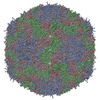
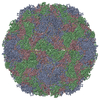
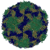
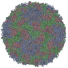
| Details | The asymmetric unit contains 20 copies of the protomer. Each protomer consists of one copy each of VP1, VP2, VP3 and VP4. The biological assembly contains 60 copies of the protomer. |
-Components
| #1: Protein | Mass: 32699.773 Da / Num. of mol.: 1 / Source method: isolated from a natural source Details: isolated in Fuyang, Anhui in 2008, grow in RD cells Source: (natural) Enterovirus A71Strain: clinical C4 strain, AH08/06, GenBank accession no. HQ611148 References: UniProt: S5QA87 |
|---|---|
| #2: Protein | Mass: 27726.135 Da / Num. of mol.: 1 / Source method: isolated from a natural source Details: isolated in Fuyang, Anhui in 2008, grow in RD cells Source: (natural) Enterovirus A71Strain: clinical C4 strain, AH08/06, GenBank accession no. HQ611148 References: UniProt: S4VM80 |
| #3: Protein | Mass: 26440.148 Da / Num. of mol.: 1 / Source method: isolated from a natural source Details: isolated in Fuyang, Anhui in 2008, grow in RD cells Source: (natural) Enterovirus A71Strain: clinical C4 strain, AH08/06, GenBank accession no. HQ611148 References: UniProt: S5ZCI0 |
| #4: Protein | Mass: 7501.162 Da / Num. of mol.: 1 / Source method: isolated from a natural source Details: isolated in Fuyang, Anhui in 2008, grow in RD cells Source: (natural) Enterovirus A71Strain: clinical C4 strain, AH08/06, GenBank accession no. HQ611148 References: UniProt: S6C3M9 |
| #5: Chemical | ChemComp-SPH /   Mass: 299.492 Da / Num. of mol.: 1 / Source method: obtained synthetically / Formula: C18H37NO2 Mass: 299.492 Da / Num. of mol.: 1 / Source method: obtained synthetically / Formula: C18H37NO2 |
-Experimental details
-Experiment
| Experiment | Method: X-RAY DIFFRACTION / Number of used crystals: 1 |
|---|
- Sample preparation
| Crystal grow | Temperature: 289 K / Method: hanging drop / pH: 4.5 Details: 0.1M Sodium Acetate containing 3.5M Sodium Formate, pH 4.5, hanging drop, temperature 289K |
|---|
-Data collection
| Diffraction | Mean temperature: 100 K | ||||||||||||||||||||||||||||||||||||||||||||||||||||||||||||||||||||||||||||||||||||||||||||||||||||||||||||||||
|---|---|---|---|---|---|---|---|---|---|---|---|---|---|---|---|---|---|---|---|---|---|---|---|---|---|---|---|---|---|---|---|---|---|---|---|---|---|---|---|---|---|---|---|---|---|---|---|---|---|---|---|---|---|---|---|---|---|---|---|---|---|---|---|---|---|---|---|---|---|---|---|---|---|---|---|---|---|---|---|---|---|---|---|---|---|---|---|---|---|---|---|---|---|---|---|---|---|---|---|---|---|---|---|---|---|---|---|---|---|---|---|---|---|
| Diffraction source | Source: SYNCHROTRON / Site: SSRF  / Beamline: BL17U / Wavelength: 0.9793 Å / Beamline: BL17U / Wavelength: 0.9793 Å | ||||||||||||||||||||||||||||||||||||||||||||||||||||||||||||||||||||||||||||||||||||||||||||||||||||||||||||||||
| Detector | Type: ADSC QUANTUM 315 / Detector: CCD / Date: Oct 10, 2011 | ||||||||||||||||||||||||||||||||||||||||||||||||||||||||||||||||||||||||||||||||||||||||||||||||||||||||||||||||
| Radiation | Protocol: SINGLE WAVELENGTH / Monochromatic (M) / Laue (L): M / Scattering type: x-ray | ||||||||||||||||||||||||||||||||||||||||||||||||||||||||||||||||||||||||||||||||||||||||||||||||||||||||||||||||
| Radiation wavelength | Wavelength: 0.9793 Å / Relative weight: 1 | ||||||||||||||||||||||||||||||||||||||||||||||||||||||||||||||||||||||||||||||||||||||||||||||||||||||||||||||||
| Reflection | Resolution: 3.306→48.286 Å / Num. all: 505463 / Num. obs: 314499 / % possible obs: 62.22 % / Observed criterion σ(F): 5 / Observed criterion σ(I): 5 / Redundancy: 1.5 % / Rmerge(I) obs: 0.216 / Χ2: 2.289 / Net I/σ(I): 4.4 | ||||||||||||||||||||||||||||||||||||||||||||||||||||||||||||||||||||||||||||||||||||||||||||||||||||||||||||||||
| Reflection shell | Diffraction-ID: 1
|
- Processing
| Software |
| ||||||||||||||||||||||||||||
|---|---|---|---|---|---|---|---|---|---|---|---|---|---|---|---|---|---|---|---|---|---|---|---|---|---|---|---|---|---|
| Refinement | Method to determine structure: MOLECULAR REPLACEMENT Starting model: PDB ENTRY 1D4M Resolution: 3.3063→48.286 Å / Occupancy max: 1 / Occupancy min: 1 / σ(F): 0 / Stereochemistry target values: Engh & Huber
| ||||||||||||||||||||||||||||
| Displacement parameters | Biso max: 116.42 Å2 / Biso mean: 50.47 Å2 / Biso min: 0 Å2 | ||||||||||||||||||||||||||||
| Refine analyze | Luzzati coordinate error obs: 0.43 Å | ||||||||||||||||||||||||||||
| Refinement step | Cycle: LAST / Resolution: 3.3063→48.286 Å
| ||||||||||||||||||||||||||||
| Refine LS restraints |
| ||||||||||||||||||||||||||||
| LS refinement shell | Resolution: 3.3063→3.389 Å
|
