 Movie
Movie Controller

[English] 日本語
 Yorodumi
- PDB-1k5m: Crystal Structure of a Human Rhinovirus Type 14:Human Immunodefic... -
+ Open data

- Basic information
| Entry | Database: PDB / ID: 1k5m | ||||||
|---|---|---|---|---|---|---|---|
| Title | Crystal Structure of a Human Rhinovirus Type 14:Human Immunodeficiency Virus Type 1 V3 Loop Chimeric Virus MN-III-2 | ||||||
 Components Components |
| ||||||
 Keywords Keywords | VIRUS / engineered rhinovirus / HIV-1 V3 loop / beta turns / Icosahedral virus | ||||||
| Function / homology |  Function and homology information Function and homology informationlysis of host organelle involved in viral entry into host cell / Dectin-2 family / symbiont-mediated suppression of host cytoplasmic pattern recognition receptor signaling pathway via inhibition of RIG-I activity / symbiont-mediated perturbation of host defense response / positive regulation of plasma membrane raft polarization / positive regulation of receptor clustering / host cell endosome membrane / picornain 2A / symbiont-mediated suppression of host mRNA export from nucleus / symbiont genome entry into host cell via pore formation in plasma membrane ...lysis of host organelle involved in viral entry into host cell / Dectin-2 family / symbiont-mediated suppression of host cytoplasmic pattern recognition receptor signaling pathway via inhibition of RIG-I activity / symbiont-mediated perturbation of host defense response / positive regulation of plasma membrane raft polarization / positive regulation of receptor clustering / host cell endosome membrane / picornain 2A / symbiont-mediated suppression of host mRNA export from nucleus / symbiont genome entry into host cell via pore formation in plasma membrane / picornain 3C / T=pseudo3 icosahedral viral capsid / host cell cytoplasmic vesicle membrane / ribonucleoside triphosphate phosphatase activity / nucleoside-triphosphate phosphatase / channel activity / monoatomic ion transmembrane transport / clathrin-dependent endocytosis of virus by host cell / DNA replication / RNA helicase activity / viral protein processing / endocytosis involved in viral entry into host cell / fusion of virus membrane with host plasma membrane / symbiont-mediated activation of host autophagy / RNA-directed RNA polymerase / cysteine-type endopeptidase activity / viral RNA genome replication / RNA-directed RNA polymerase activity / fusion of virus membrane with host endosome membrane / viral envelope / virion attachment to host cell / DNA-templated transcription / host cell nucleus / host cell plasma membrane / virion membrane / structural molecule activity / proteolysis / RNA binding / zinc ion binding / ATP binding / membrane / identical protein binding Similarity search - Function | ||||||
| Biological species |  Human rhinovirus 14Human immunodeficiency virus type 1 group M subtype B Human rhinovirus 14Human immunodeficiency virus type 1 group M subtype B | ||||||
| Method |  X-RAY DIFFRACTION / SYNCHROTRON / MOLECULAR REPLACEMENT / Resolution: 2.7 Å X-RAY DIFFRACTION / SYNCHROTRON / MOLECULAR REPLACEMENT / Resolution: 2.7 Å | ||||||
 Authors Authors | Ding, J. / Smith, A.D. / Geisler, S.C. / Ma, X. / Arnold, G.F. / Arnold, E. | ||||||
 Citation Citation | Journal: Structure / Year: 2002 Title: Crystal Structure of a Human Rhinovirus that Displays Part of the HIV-1 V3 Loop and Induces Neutralizing Antibodies against HIV-1 Authors: Ding, J. / Smith, A.D. / Geisler, S.C. / Ma, X. / Arnold, G.F. / Arnold, E. #1: Journal: J.Virol. / Year: 1998Title: Human rhinovirus type 14:human immunodeficiency virus type 1 (HIV-1) V3 loop chimeras from a combinational library induce potent neutralizing antibody responses against HIV-1 Authors: Smith, A.D. / Geisler, S.C. / Chen, A.A. / Resnick, D.A. / Roy, B.M. / Lewi, P.J. / Arnold, E. / Arnold, G.F. | ||||||
| History |
|
- Structure visualization
| Structure viewer | Molecule: MolmilJmol/JSmol |
|---|
- Downloads & links
-Download
| PDBx/mmCIF format | 1k5m.cif.gz | 189.4 KB | Display | PDBx/mmCIF format |
|---|---|---|---|---|
| PDB format | pdb1k5m.ent.gz | 145.7 KB | Display | PDB format |
| PDBx/mmJSON format | 1k5m.json.gz | Tree view | PDBx/mmJSON format | |
| Others |  Other downloads Other downloads |
-Validation report
| Arichive directory | https://data.pdbj.org/pub/pdb/validation_reports/k5/1k5mftp://data.pdbj.org/pub/pdb/validation_reports/k5/1k5m | HTTPS FTP |
|---|
-Related structure data
| Related structure data |  4rhvS S: Starting model for refinement |
|---|---|
| Similar structure data |


-Links
 PDBj
PDBj


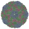
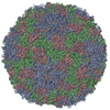
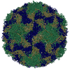
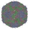
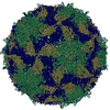
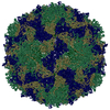
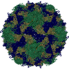
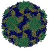
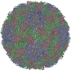
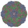
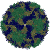
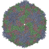
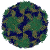
- Assembly
| Deposited unit | 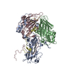
| ||||||||||||||||||||||||||||||||||||||||||||||||
|---|---|---|---|---|---|---|---|---|---|---|---|---|---|---|---|---|---|---|---|---|---|---|---|---|---|---|---|---|---|---|---|---|---|---|---|---|---|---|---|---|---|---|---|---|---|---|---|---|---|
| 1 | x 60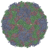
| ||||||||||||||||||||||||||||||||||||||||||||||||
| 2 |
| ||||||||||||||||||||||||||||||||||||||||||||||||
| 3 | x 5
| ||||||||||||||||||||||||||||||||||||||||||||||||
| 4 | x 6
| ||||||||||||||||||||||||||||||||||||||||||||||||
| 5 | 
| ||||||||||||||||||||||||||||||||||||||||||||||||
| 6 | x 15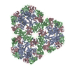
| ||||||||||||||||||||||||||||||||||||||||||||||||
| Unit cell |
| ||||||||||||||||||||||||||||||||||||||||||||||||
| Symmetry | Point symmetry: (Hermann–Mauguin notation: 532 / Schoenflies symbol: I (icosahedral)) | ||||||||||||||||||||||||||||||||||||||||||||||||
| Noncrystallographic symmetry (NCS) | NCS oper:
|
-Components
-COAT PROTEIN ... , 3 types, 3 molecules ACD
| #1: Protein | Mass: 32560.549 Da / Num. of mol.: 1 Source method: isolated from a genetically manipulated source Source: (gene. exp.) Human rhinovirus 14 / Genus: Rhinovirus / Species: Human rhinovirus B / Plasmid: p3IIST-MN-III-2 / Cell line (production host): H1-HeLa cells / Production host:  Homo sapiens (human) / References: UniProt: P03303 Homo sapiens (human) / References: UniProt: P03303 |
|---|---|
| #3: Protein | Mass: 26236.754 Da / Num. of mol.: 1 Source method: isolated from a genetically manipulated source Source: (gene. exp.) Human rhinovirus 14 / Genus: Rhinovirus / Species: Human rhinovirus B / Plasmid: p3IIST-MN-III-2 / Cell line (production host): H1-HeLa cells / Production host: Homo sapiens (human) / References: UniProt: P03303 |
| #4: Protein | Mass: 7183.863 Da / Num. of mol.: 1 Source method: isolated from a genetically manipulated source Source: (gene. exp.) Human rhinovirus 14 / Genus: Rhinovirus / Species: Human rhinovirus B / Plasmid: p3IIST-MN-III-2 / Cell line (production host): H1-HeLa cells / Production host: Homo sapiens (human) / References: UniProt: P03303 |
-Protein , 1 types, 1 molecules B
| #2: Protein | Mass: 30097.109 Da / Num. of mol.: 1 Source method: isolated from a genetically manipulated source Details: THE CHIMERA CONSISTS OF THE HRV14 COAT PROTEIN VP2 (P1B) AND RESIDUES 314-325 OF HIV-1 gp120. Source: (gene. exp.) Human rhinovirus 14, (gene. exp.) Human immunodeficiency virus type 1 group M subtype B (isolate MN)Genus: Rhinovirus / Species: Human rhinovirus B / Plasmid: p3IIST-MN-III-2 / Gene: env / Cell line (production host): H1-HeLa cells / Production host: Homo sapiens (human) / References: UniProt: P03303, UniProt: P05877 |
|---|
-Non-polymers , 2 types, 638 molecules 

| #5: Chemical | ChemComp-SPH /  Mass: 299.492 Da / Num. of mol.: 1 / Source method: obtained synthetically / Formula: C18H37NO2 Mass: 299.492 Da / Num. of mol.: 1 / Source method: obtained synthetically / Formula: C18H37NO2 |
|---|---|
| #6: Water | ChemComp-HOH / Mass: 18.015 Da / Num. of mol.: 637 / Source method: isolated from a natural source / Formula: H2O |
-Experimental details
-Experiment
| Experiment | Method: X-RAY DIFFRACTION / Number of used crystals: 2 |
|---|
- Sample preparation
| Crystal | Density Matthews: 3.75 Å3/Da / Density % sol: 67.2 % | ||||||||||||||||||
|---|---|---|---|---|---|---|---|---|---|---|---|---|---|---|---|---|---|---|---|
| Crystal grow | Temperature: 298 K / Method: vapor diffusion, hanging drop / pH: 7.5 Details: 1.5 M ammonium formate and 0.15 M sodium HEPES, pH 7.5, VAPOR DIFFUSION, HANGING DROP, temperature 298K | ||||||||||||||||||
| Crystal grow | *PLUS | ||||||||||||||||||
| Components of the solutions | *PLUS
|
-Data collection
| Diffraction |
| ||||||||||||||||||
|---|---|---|---|---|---|---|---|---|---|---|---|---|---|---|---|---|---|---|---|
| Diffraction source |
| ||||||||||||||||||
| Detector |
| ||||||||||||||||||
| Radiation |
| ||||||||||||||||||
| Radiation wavelength |
| ||||||||||||||||||
| Reflection | Resolution: 2.7→50 Å / Num. all: 513239 / Num. obs: 513239 / % possible obs: 92.8 % / Observed criterion σ(F): 0 / Observed criterion σ(I): 0 / Redundancy: 13.8 % / Biso Wilson estimate: 29 Å2 / Rmerge(I) obs: 0.114 / Net I/σ(I): 10 | ||||||||||||||||||
| Reflection shell | Resolution: 2.7→2.8 Å / Redundancy: 5 % / Rmerge(I) obs: 0.212 / Mean I/σ(I) obs: 4 / Num. unique all: 46826 / % possible all: 82.7 | ||||||||||||||||||
| Reflection | *PLUS Lowest resolution: 50 Å / % possible obs: 92.4 % / Num. measured all: 7100958 | ||||||||||||||||||
| Reflection shell | *PLUS % possible obs: 81.6 % |

- Processing
| Software |
| |||||||||||||||||||||||||||||||||||
|---|---|---|---|---|---|---|---|---|---|---|---|---|---|---|---|---|---|---|---|---|---|---|---|---|---|---|---|---|---|---|---|---|---|---|---|---|
| Refinement | Method to determine structure: MOLECULAR REPLACEMENT Starting model: Wild-type HRV14 structure coordinates (PDB entry 4RHV) Resolution: 2.7→50 Å / Isotropic thermal model: isotropic Cross valid method: The free R-factor was calculated in early stages of model building and refinement to monitor the progress. Due to the strong interdependency of structure factors in the presence ...Cross valid method: The free R-factor was calculated in early stages of model building and refinement to monitor the progress. Due to the strong interdependency of structure factors in the presence of high NCS, the free R-factor did not provide any useful information. Therefore, in the later stages of structure refinement, all data were included and no free R factor was calculated. σ(F): 2 / Stereochemistry target values: Engh & Huber
| |||||||||||||||||||||||||||||||||||
| Displacement parameters | Biso mean: 18.4 Å2 | |||||||||||||||||||||||||||||||||||
| Refine analyze | Luzzati coordinate error obs: 0.25 Å | |||||||||||||||||||||||||||||||||||
| Refinement step | Cycle: LAST / Resolution: 2.7→50 Å
| |||||||||||||||||||||||||||||||||||
| Refine LS restraints |
| |||||||||||||||||||||||||||||||||||
| LS refinement shell | Refine-ID: X-RAY DIFFRACTION / Num. reflection Rfree: _ / Total num. of bins used: 10
| |||||||||||||||||||||||||||||||||||
| Xplor file | Serial no: 1 / Param file: parhcsdx.pro / Topol file: tophcsdx.pro | |||||||||||||||||||||||||||||||||||
| Refinement | *PLUS Lowest resolution: 50 Å / σ(F): 2 / Rfactor all: 0.22 | |||||||||||||||||||||||||||||||||||
| Solvent computation | *PLUS | |||||||||||||||||||||||||||||||||||
| Displacement parameters | *PLUS | |||||||||||||||||||||||||||||||||||
| Refine LS restraints | *PLUS
| |||||||||||||||||||||||||||||||||||
| LS refinement shell | *PLUS Highest resolution: 2.7 Å / Lowest resolution: 2.8 Å / Rfactor all: 0.287 |
