 Movie
Movie Controller


+ Open data

- Basic information
| Entry | Database: PDB / ID: 4qpg | ||||||
|---|---|---|---|---|---|---|---|
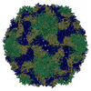
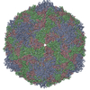
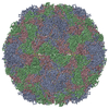
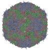
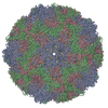
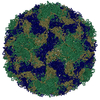
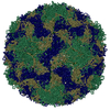
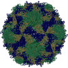
| Title | Crystal structure of empty hepatitis A virus | ||||||
 Components Components |
| ||||||
 Keywords Keywords | VIRUS / evolution / insect picorna-like viruses / ICOSAHEDRAL / domain swap / beta-barrel / PICORNAVIRUS / pathogen / Liver | ||||||
| Function / homology |  Function and homology information Function and homology informationhost cell mitochondrial outer membrane / host cell membrane / symbiont-mediated suppression of host cytoplasmic pattern recognition receptor signaling pathway via inhibition of MAVS activity / picornain 3C / T=pseudo3 icosahedral viral capsid / host cell cytoplasmic vesicle membrane / host multivesicular body / ribonucleoside triphosphate phosphatase activity / nucleoside-triphosphate phosphatase / channel activity ...host cell mitochondrial outer membrane / host cell membrane / symbiont-mediated suppression of host cytoplasmic pattern recognition receptor signaling pathway via inhibition of MAVS activity / picornain 3C / T=pseudo3 icosahedral viral capsid / host cell cytoplasmic vesicle membrane / host multivesicular body / ribonucleoside triphosphate phosphatase activity / nucleoside-triphosphate phosphatase / channel activity / monoatomic ion transmembrane transport / RNA helicase activity / RNA-directed RNA polymerase / cysteine-type endopeptidase activity / viral RNA genome replication / RNA-directed RNA polymerase activity / symbiont entry into host cell / virion attachment to host cell / DNA-templated transcription / structural molecule activity / proteolysis / RNA binding / ATP binding Similarity search - Function | ||||||
| Biological species |  Human hepatitis A virus Human hepatitis A virus | ||||||
| Method |  X-RAY DIFFRACTION / SYNCHROTRON / MOLECULAR REPLACEMENT / Resolution: 3.5 Å X-RAY DIFFRACTION / SYNCHROTRON / MOLECULAR REPLACEMENT / Resolution: 3.5 Å | ||||||
 Authors Authors | Wang, X. / Ren, J. / Gao, Q. / Hu, Z. / Sun, Y. / Li, X. / Rowlands, D.J. / Yin, W. / Wang, J. / Stuart, D.I. ...Wang, X. / Ren, J. / Gao, Q. / Hu, Z. / Sun, Y. / Li, X. / Rowlands, D.J. / Yin, W. / Wang, J. / Stuart, D.I. / Rao, Z. / Fry, E.E. | ||||||
 Citation Citation | Journal: Nature / Year: 2015 Title: Hepatitis A virus and the origins of picornaviruses. Authors: Wang, X. / Ren, J. / Gao, Q. / Hu, Z. / Sun, Y. / Li, X. / Rowlands, D.J. / Yin, W. / Wang, J. / Stuart, D.I. / Rao, Z. / Fry, E.E. | ||||||
| History |
|
- Structure visualization
| Structure viewer | Molecule: MolmilJmol/JSmol |
|---|
- Downloads & links
-Download
| PDBx/mmCIF format | 4qpg.cif.gz | 151.5 KB | Display | PDBx/mmCIF format |
|---|---|---|---|---|
| PDB format | pdb4qpg.ent.gz | 117.9 KB | Display | PDB format |
| PDBx/mmJSON format | 4qpg.json.gz | Tree view | PDBx/mmJSON format | |
| Others |  Other downloads Other downloads |
-Validation report
| Arichive directory | https://data.pdbj.org/pub/pdb/validation_reports/qp/4qpgftp://data.pdbj.org/pub/pdb/validation_reports/qp/4qpg | HTTPS FTP |
|---|
-Related structure data
| Related structure data | 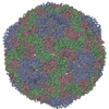 4qpiC  1fmdS S: Starting model for refinement C: citing same article ( |
|---|---|
| Similar structure data |


-Links
 PDBj
PDBj

- Assembly
| Deposited unit | 
| |||||||||||||||||||||||||||||||||||||||||||||||||||||||||||||||||||||||||||||||||||||||||||||||||||||||||||||||||||
|---|---|---|---|---|---|---|---|---|---|---|---|---|---|---|---|---|---|---|---|---|---|---|---|---|---|---|---|---|---|---|---|---|---|---|---|---|---|---|---|---|---|---|---|---|---|---|---|---|---|---|---|---|---|---|---|---|---|---|---|---|---|---|---|---|---|---|---|---|---|---|---|---|---|---|---|---|---|---|---|---|---|---|---|---|---|---|---|---|---|---|---|---|---|---|---|---|---|---|---|---|---|---|---|---|---|---|---|---|---|---|---|---|---|---|---|---|
| 1 | x 60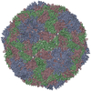
| |||||||||||||||||||||||||||||||||||||||||||||||||||||||||||||||||||||||||||||||||||||||||||||||||||||||||||||||||||
| 2 |
| |||||||||||||||||||||||||||||||||||||||||||||||||||||||||||||||||||||||||||||||||||||||||||||||||||||||||||||||||||
| 3 | x 5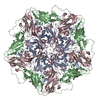
| |||||||||||||||||||||||||||||||||||||||||||||||||||||||||||||||||||||||||||||||||||||||||||||||||||||||||||||||||||
| 4 | x 6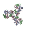
| |||||||||||||||||||||||||||||||||||||||||||||||||||||||||||||||||||||||||||||||||||||||||||||||||||||||||||||||||||
| 5 | 
| |||||||||||||||||||||||||||||||||||||||||||||||||||||||||||||||||||||||||||||||||||||||||||||||||||||||||||||||||||
| 6 | x 30
| |||||||||||||||||||||||||||||||||||||||||||||||||||||||||||||||||||||||||||||||||||||||||||||||||||||||||||||||||||
| Unit cell |
| |||||||||||||||||||||||||||||||||||||||||||||||||||||||||||||||||||||||||||||||||||||||||||||||||||||||||||||||||||
| Symmetry | Point symmetry: (Schoenflies symbol: I (icosahedral)) | |||||||||||||||||||||||||||||||||||||||||||||||||||||||||||||||||||||||||||||||||||||||||||||||||||||||||||||||||||
| Noncrystallographic symmetry (NCS) | NCS oper:
|