 ムービー
ムービー コントローラー


+ データを開く

- 基本情報
基本情報
| 登録情報 | データベース: PDB / ID: 1ovt | ||||||
|---|---|---|---|---|---|---|---|
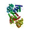
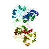
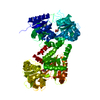
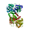
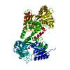
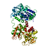
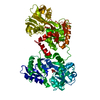
| タイトル | REFINED CRYSTALLOGRAPHIC STRUCTURE OF HEN OVOTRANSFERRIN AT 2.4 ANGSTROMS RESOLUTION | ||||||
 要素 要素 | OVOTRANSFERRIN | ||||||
 キーワード キーワード | IRON TRANSPORT PROTEIN | ||||||
| 機能・相同性 |  機能・相同性情報 機能・相同性情報organomineral extracellular matrix / iron ion transmembrane transport / antimicrobial humoral response / ferric iron binding / acute-phase response / iron ion transport / recycling endosome / antibacterial humoral response / response to lipopolysaccharide / intracellular iron ion homeostasis ...organomineral extracellular matrix / iron ion transmembrane transport / antimicrobial humoral response / ferric iron binding / acute-phase response / iron ion transport / recycling endosome / antibacterial humoral response / response to lipopolysaccharide / intracellular iron ion homeostasis / early endosome / iron ion binding / response to xenobiotic stimulus / : / plasma membrane 類似検索 - 分子機能 | ||||||
| 生物種 |  | ||||||
| 手法 |  X線回折 / 解像度: 2.4 Å X線回折 / 解像度: 2.4 Å | ||||||
 データ登録者 データ登録者 | Kurokawa, H. / Mikami, B. / Hirose, M. | ||||||
 引用 引用 | ジャーナル: J.Mol.Biol. / 年: 1995 タイトル: Crystal structure of diferric hen ovotransferrin at 2.4 A resolution. 著者: Kurokawa, H. / Mikami, B. / Hirose, M. #1: ジャーナル: Biochemistry / 年: 1993タイトル: Structural Evidence for a Ph Sensitive Di-Lysine Trigger in the Hen Ovotransferrin N-Lobe: Implications for Transferrin Iron Release 著者: Dewan, J.C. / Mikami, B. / Hirose, M. / Sacchettini, J.C. #2: ジャーナル: J.Biochem.(Tokyo) / 年: 1990タイトル: Crystallization of N-Terminal Lobe of Ovotransferrin 著者: Mikami, B. / Hirose, M. | ||||||
| 履歴 |
| ||||||
| Remark 700 | SHEET THERE IS ONE BIFURCATED SHEET IN EACH OF THE TWO LOBES OF OVOTRANSFERRIN. SHEET S1 IS ...SHEET THERE IS ONE BIFURCATED SHEET IN EACH OF THE TWO LOBES OF OVOTRANSFERRIN. SHEET S1 IS PRESENTED AS SHEETS S1A, S1B, AND S1C ON SHEET RECORDS BELOW. NOTE THAT STRANDS 4 OF S1A AND 3 OF S1B ARE IDENTICAL, STRANDS 1 OF S1B AND 3 OF S1C ARE IDENTICAL, AND STRANDS 2 OF S1B AND 4 OF S1C ARE IDENTICAL. SHEET S2 IS PRESENTED AS SHEETS S2A, S2B, AND S2C ON SHEET RECORDS BELOW. NOTE THAT STRANDS 4 OF S2A AND 3 OF S2B ARE IDENTICAL, STRANDS 1 OF S2B AND 3 OF S2C ARE IDENTICAL, AND STRANDS 2 OF S2B AND 4 OF S2C ARE IDENTICAL. |
- 構造の表示
構造の表示
| 構造ビューア | 分子: MolmilJmol/JSmol |
|---|
- ダウンロードとリンク
-ダウンロード
| PDBx/mmCIF形式 | 1ovt.cif.gz | 146.4 KB | 表示 | PDBx/mmCIF形式 |
|---|---|---|---|---|
| PDB形式 | pdb1ovt.ent.gz | 113.8 KB | 表示 | PDB形式 |
| PDBx/mmJSON形式 | 1ovt.json.gz | ツリー表示 | PDBx/mmJSON形式 | |
| その他 |  その他のダウンロード その他のダウンロード |
-検証レポート
| アーカイブディレクトリ | https://data.pdbj.org/pub/pdb/validation_reports/ov/1ovtftp://data.pdbj.org/pub/pdb/validation_reports/ov/1ovt | HTTPS FTP |
|---|
-関連構造データ


-リンク
 PDBj
PDBj


- 集合体
集合体
| 登録構造単位 | 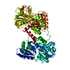
| ||||||||
|---|---|---|---|---|---|---|---|---|---|
| 1 |
| ||||||||
| 単位格子 |
| ||||||||
| Atom site foot note | 1: CIS PROLINE - PRO 71 / 2: CIS PROLINE - PRO 287 3: SER 339 - PRO 340 OMEGA = 143.08 PEPTIDE BOND DEVIATES SIGNIFICANTLY FROM TRANS CONFORMATION 4: ARG 427 - PRO 428 OMEGA = 238.29 PEPTIDE BOND DEVIATES SIGNIFICANTLY FROM TRANS CONFORMATION 5: PRO 509 - PRO 510 OMEGA = 250.41 PEPTIDE BOND DEVIATES SIGNIFICANTLY FROM TRANS CONFORMATION |
-要素
| #1: タンパク質 | 分子量: 75929.008 Da / 分子数: 1 / 由来タイプ: 天然 / 詳細: DIFERRIC FORM / 由来: (天然) | ||||||
|---|---|---|---|---|---|---|---|
| #2: 化合物 |   分子量: 55.845 Da / 分子数: 2 / 由来タイプ: 合成 / 式: Fe 分子量: 55.845 Da / 分子数: 2 / 由来タイプ: 合成 / 式: Fe#3: 化合物 |   分子量: 60.009 Da / 分子数: 2 / 由来タイプ: 合成 / 式: CO3 分子量: 60.009 Da / 分子数: 2 / 由来タイプ: 合成 / 式: CO3#4: 水 | ChemComp-HOH / |  分子量: 18.015 Da / 分子数: 132 / 由来タイプ: 天然 / 式: H2O 分子量: 18.015 Da / 分子数: 132 / 由来タイプ: 天然 / 式: H2OHas protein modification | Y | |
-実験情報
-実験
| 実験 | 手法: X線回折 |
|---|
- 試料調製
試料調製
| 結晶 | マシュー密度: 2.67 Å3/Da / 溶媒含有率: 53.94 % | ||||||||||||||||||||
|---|---|---|---|---|---|---|---|---|---|---|---|---|---|---|---|---|---|---|---|---|---|
| 結晶化 | *PLUS 温度: 4 ℃ / pH: 5.9 / 手法: batch method | ||||||||||||||||||||
| 溶液の組成 | *PLUS
|
-データ収集
| 検出器 | 日付: 1993年12月3日 |
|---|---|
| 放射 | 単色(M)・ラウエ(L): M / 散乱光タイプ: x-ray |
| 放射波長 | 相対比: 1 |
| 反射 | Num. obs: 28473 / % possible obs: 87 % / 冗長度: 3.2 % |
| 反射 | *PLUS 最高解像度: 2.4 Å / 最低解像度: 11 Å / Observed criterion σ(F): 2 / Num. measured all: 61204 |
- 解析
解析
| ソフトウェア |
| ||||||||||||||||||||||||||||||||||||||||||||||||||||||||||||
|---|---|---|---|---|---|---|---|---|---|---|---|---|---|---|---|---|---|---|---|---|---|---|---|---|---|---|---|---|---|---|---|---|---|---|---|---|---|---|---|---|---|---|---|---|---|---|---|---|---|---|---|---|---|---|---|---|---|---|---|---|---|
| 精密化 | 解像度: 2.4→11 Å / σ(F): 2 /
| ||||||||||||||||||||||||||||||||||||||||||||||||||||||||||||
| 原子変位パラメータ | Biso mean: 24 Å2 | ||||||||||||||||||||||||||||||||||||||||||||||||||||||||||||
| Refine analyze | Luzzati coordinate error obs: 0.25 Å | ||||||||||||||||||||||||||||||||||||||||||||||||||||||||||||
| 精密化ステップ | サイクル: LAST / 解像度: 2.4→11 Å
| ||||||||||||||||||||||||||||||||||||||||||||||||||||||||||||
| 拘束条件 |
| ||||||||||||||||||||||||||||||||||||||||||||||||||||||||||||
| ソフトウェア | *PLUS 名称: X-PLOR / 分類: refinement | ||||||||||||||||||||||||||||||||||||||||||||||||||||||||||||
| 精密化 | *PLUS Rfactor Rfree: 0.265 | ||||||||||||||||||||||||||||||||||||||||||||||||||||||||||||
| 溶媒の処理 | *PLUS | ||||||||||||||||||||||||||||||||||||||||||||||||||||||||||||
| 原子変位パラメータ | *PLUS | ||||||||||||||||||||||||||||||||||||||||||||||||||||||||||||
| 拘束条件 | *PLUS
|
