 Movie
Movie Controller


+ Open data

- Basic information
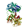
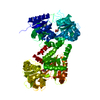
| Entry | Database: PDB / ID: 1b0l | ||||||
|---|---|---|---|---|---|---|---|
| Title | RECOMBINANT HUMAN DIFERRIC LACTOFERRIN | ||||||
 Components Components | PROTEIN (LACTOFERRIN) | ||||||
 Keywords Keywords | METAL BINDING PROTEIN / TRANSFERRIN / BINDING PROTEIN / METALLOPROTEIN | ||||||
| Function / homology |  Function and homology information Function and homology informationmembrane destabilizing activity / host-mediated suppression of viral proces / Mtb iron assimilation by chelation / phagocytic vesicle lumen / Metal sequestration by antimicrobial proteins / negative regulation of viral process / negative regulation of tumor necrosis factor (ligand) superfamily member 11 production / positive regulation of toll-like receptor 4 signaling pathway / negative regulation of single-species biofilm formation in or on host organism / positive regulation of bone mineralization involved in bone maturation ...membrane destabilizing activity / host-mediated suppression of viral proces / Mtb iron assimilation by chelation / phagocytic vesicle lumen / Metal sequestration by antimicrobial proteins / negative regulation of viral process / negative regulation of tumor necrosis factor (ligand) superfamily member 11 production / positive regulation of toll-like receptor 4 signaling pathway / negative regulation of single-species biofilm formation in or on host organism / positive regulation of bone mineralization involved in bone maturation / negative regulation of ATP-dependent activity / negative regulation of osteoclast development / specific granule / negative regulation of lipopolysaccharide-mediated signaling pathway / positive regulation of chondrocyte proliferation / antifungal humoral response / regulation of tumor necrosis factor production / bone morphogenesis / Antimicrobial peptides / positive regulation of osteoblast proliferation / negative regulation of viral genome replication / positive regulation of protein serine/threonine kinase activity / humoral immune response / Hydrolases; Acting on peptide bonds (peptidases); Serine endopeptidases / positive regulation of osteoblast differentiation / cysteine-type endopeptidase inhibitor activity / : / regulation of cytokine production / secretory granule / ossification / protein serine/threonine kinase activator activity / innate immune response in mucosa / iron ion transport / lipopolysaccharide binding / recycling endosome / specific granule lumen / tertiary granule lumen / antimicrobial humoral immune response mediated by antimicrobial peptide / heparin binding / antibacterial humoral response / killing of cells of another organism / defense response to Gram-negative bacterium / early endosome / positive regulation of canonical NF-kappaB signal transduction / iron ion binding / Amyloid fiber formation / serine-type endopeptidase activity / Neutrophil degranulation / negative regulation of apoptotic process / cell surface / protein-containing complex / proteolysis / : / DNA binding / extracellular exosome / extracellular region / nucleus / plasma membrane / cytoplasm Similarity search - Function | ||||||
| Biological species |  Homo sapiens (human) Homo sapiens (human) | ||||||
| Method |  X-RAY DIFFRACTION / OTHER / Resolution: 2.2 Å X-RAY DIFFRACTION / OTHER / Resolution: 2.2 Å | ||||||
 Authors Authors | Baker, E.N. / Jameson, G.B. / Sun, X. | ||||||
 Citation Citation | Journal: Acta Crystallogr.,Sect.D / Year: 1999 Title: Structure of recombinant human lactoferrin expressed in Aspergillus awamori. Authors: Sun, X.L. / Baker, H.M. / Shewry, S.C. / Jameson, G.B. / Baker, E.N. #1: Journal: Acta Crystallogr.,Sect.D / Year: 1995Title: Structure of Human Diferric Lactoferrin Refined at 2.2 A Resolution Authors: Haridas, M. / Anderson, B.F. / Baker, E.N. | ||||||
| History |
| ||||||
| Remark 650 | HELIX DETERMINATION METHOD: AUTHOR-DETERMINED |
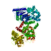
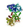
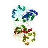
- Structure visualization
| Structure viewer | Molecule: MolmilJmol/JSmol |
|---|
- Downloads & links
-Download
| PDBx/mmCIF format | 1b0l.cif.gz | 150.8 KB | Display | PDBx/mmCIF format |
|---|---|---|---|---|
| PDB format | pdb1b0l.ent.gz | 117.1 KB | Display | PDB format |
| PDBx/mmJSON format | 1b0l.json.gz | Tree view | PDBx/mmJSON format | |
| Others |  Other downloads Other downloads |
-Validation report
| Arichive directory | https://data.pdbj.org/pub/pdb/validation_reports/b0/1b0lftp://data.pdbj.org/pub/pdb/validation_reports/b0/1b0l | HTTPS FTP |
|---|
-Related structure data
| Related structure data | 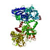 1lfgS S: Starting model for refinement |
|---|---|
| Similar structure data |

-Links
 PDBj
PDBj


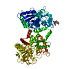
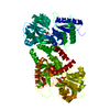
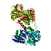
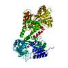
- Assembly
| Deposited unit | 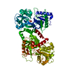
| ||||||||
|---|---|---|---|---|---|---|---|---|---|
| 1 |
| ||||||||
| Unit cell |
|
-Components
| #1: Protein | Mass: 76293.289 Da / Num. of mol.: 1 / Mutation: A10T Source method: isolated from a genetically manipulated source Source: (gene. exp.) Homo sapiens (human) / Production host:  | ||||||||
|---|---|---|---|---|---|---|---|---|---|
| #2: Chemical |   Mass: 55.845 Da / Num. of mol.: 2 / Source method: obtained synthetically / Formula: Fe Mass: 55.845 Da / Num. of mol.: 2 / Source method: obtained synthetically / Formula: Fe#3: Chemical |   Mass: 60.009 Da / Num. of mol.: 2 / Source method: obtained synthetically / Formula: CO3 Mass: 60.009 Da / Num. of mol.: 2 / Source method: obtained synthetically / Formula: CO3#4: Water | ChemComp-HOH / |  Mass: 18.015 Da / Num. of mol.: 294 / Source method: isolated from a natural source / Formula: H2O Mass: 18.015 Da / Num. of mol.: 294 / Source method: isolated from a natural source / Formula: H2OCompound details | TWO-FOLD INTERNAL SEQUENCE HOMOLOGY (~ 40% IDENTITY). ONE BINDING SITE IN EACH LOBE. | Has protein modification | Y | |
-Experimental details
-Experiment
| Experiment | Method: X-RAY DIFFRACTION / Number of used crystals: 1 |
|---|
- Sample preparation
| Crystal | Density Matthews: 2.73 Å3/Da / Density % sol: 55.89 % | ||||||||||||||||||||||||||||||||||||
|---|---|---|---|---|---|---|---|---|---|---|---|---|---|---|---|---|---|---|---|---|---|---|---|---|---|---|---|---|---|---|---|---|---|---|---|---|---|
| Crystal grow | pH: 8 Details: DIALYSIS AGAINST 10MM SODIUM PHOSPHATE PH 8.0, CONTAINING 10% ETHANOL | ||||||||||||||||||||||||||||||||||||
| Crystal grow | *PLUS Temperature: 277 K / Method: microdialysis | ||||||||||||||||||||||||||||||||||||
| Components of the solutions | *PLUS
|
-Data collection
| Diffraction | Mean temperature: 293 K |
|---|---|
| Diffraction source | Source: ROTATING ANODE / Type: RIGAKU RU200 / Wavelength: 1.5418 |
| Detector | Type: RIGAKU / Detector: IMAGE PLATE / Date: May 1, 1996 |
| Radiation | Monochromator: GRAPHITE / Protocol: SINGLE WAVELENGTH / Monochromatic (M) / Laue (L): M / Scattering type: x-ray |
| Radiation wavelength | Wavelength: 1.5418 Å / Relative weight: 1 |
| Reflection | Resolution: 2.2→40 Å / Num. obs: 36329 / % possible obs: 82.3 % / Redundancy: 2.8 % / Rmerge(I) obs: 0.046 / Net I/σ(I): 11.1 |
| Reflection shell | Resolution: 2.2→2.3 Å / Redundancy: 3.1 % / Rmerge(I) obs: 0.542 / Mean I/σ(I) obs: 1.5 / % possible all: 67.8 |
- Processing
| Software |
| |||||||||||||||||||||||||||||||||
|---|---|---|---|---|---|---|---|---|---|---|---|---|---|---|---|---|---|---|---|---|---|---|---|---|---|---|---|---|---|---|---|---|---|---|
| Refinement | Method to determine structure: OTHER Starting model: PDB ENTRY 1LFG Resolution: 2.2→10 Å / Cross valid method: THROUGHOUT / σ(F): 0 / Stereochemistry target values: ENGH AND HUBER
| |||||||||||||||||||||||||||||||||
| Refinement step | Cycle: LAST / Resolution: 2.2→10 Å
| |||||||||||||||||||||||||||||||||
| Refine LS restraints |
|
