 Movie
Movie Controller

[English] 日本語
 Yorodumi
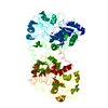
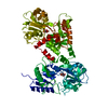
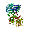
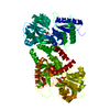
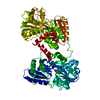
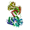
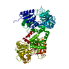
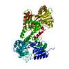
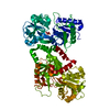
- PDB-1f9b: MELANIN PROTEIN INTERACTION: X-RAY STRUCTURE OF THE COMPLEX OF MA... -
+ Open data

- Basic information
| Entry | Database: PDB / ID: 1f9b | ||||||
|---|---|---|---|---|---|---|---|
| Title | MELANIN PROTEIN INTERACTION: X-RAY STRUCTURE OF THE COMPLEX OF MARE LACTOFERRIN WITH MELANIN MONOMERS | ||||||
 Components Components | LACTOTRANSFERRIN | ||||||
 Keywords Keywords | METAL TRANSPORT / Lactoferrin / IDQ molecule / complex / melanin / IRON TRANSPORT / METAL-BINDING | ||||||
| Function / homology |  Function and homology information Function and homology informationnegative regulation of tumor necrosis factor (ligand) superfamily member 11 production / negative regulation of single-species biofilm formation in or on host organism / positive regulation of bone mineralization involved in bone maturation / negative regulation of osteoclast development / specific granule / negative regulation of lipopolysaccharide-mediated signaling pathway / positive regulation of chondrocyte proliferation / antifungal humoral response / regulation of tumor necrosis factor production / bone morphogenesis ...negative regulation of tumor necrosis factor (ligand) superfamily member 11 production / negative regulation of single-species biofilm formation in or on host organism / positive regulation of bone mineralization involved in bone maturation / negative regulation of osteoclast development / specific granule / negative regulation of lipopolysaccharide-mediated signaling pathway / positive regulation of chondrocyte proliferation / antifungal humoral response / regulation of tumor necrosis factor production / bone morphogenesis / positive regulation of osteoblast proliferation / Hydrolases; Acting on peptide bonds (peptidases); Serine endopeptidases / positive regulation of osteoblast differentiation / regulation of cytokine production / serine-type peptidase activity / ossification / innate immune response in mucosa / iron ion transport / recycling endosome / antibacterial humoral response / early endosome / negative regulation of apoptotic process / proteolysis / : / metal ion binding / plasma membrane Similarity search - Function | ||||||
| Biological species |  | ||||||
| Method |  X-RAY DIFFRACTION / Resolution: 2.7 Å X-RAY DIFFRACTION / Resolution: 2.7 Å | ||||||
 Authors Authors | Kumar, S. / Singh, T.P. / Sharma, A.K. / Singh, N. / Raman, G. | ||||||
 Citation Citation | Journal: Proteins / Year: 2001 Title: Lactoferrin-melanin interaction and its possible implications in melanin polymerization: crystal structure of the complex formed between mare lactoferrin and melanin monomers at 2.7-A resolution. Authors: Sharma, A.K. / Kumar, S. / Sharma, V. / Nagpal, A. / Singh, N. / Tamboli, I. / Mani, I. / Raman, G. / Singh, T.P. #1: Journal: Indian J.Physics / Year: 2000Title: Metal Substitution in Lactoferrins: the Crystal Structure of Manganese Lactoferrin at 3.4 A | ||||||
| History |
|
- Structure visualization
| Structure viewer | Molecule: MolmilJmol/JSmol |
|---|
- Downloads & links
-Download
| PDBx/mmCIF format | 1f9b.cif.gz | 143.4 KB | Display | PDBx/mmCIF format |
|---|---|---|---|---|
| PDB format | pdb1f9b.ent.gz | 110.6 KB | Display | PDB format |
| PDBx/mmJSON format | 1f9b.json.gz | Tree view | PDBx/mmJSON format | |
| Others |  Other downloads Other downloads |
-Validation report
| Arichive directory | https://data.pdbj.org/pub/pdb/validation_reports/f9/1f9bftp://data.pdbj.org/pub/pdb/validation_reports/f9/1f9b | HTTPS FTP |
|---|
-Related structure data
| Related structure data | |
|---|---|
| Similar structure data |


-Links
 PDBj
PDBj


- Assembly
| Deposited unit | 
| ||||||||
|---|---|---|---|---|---|---|---|---|---|
| 1 |
| ||||||||
| Unit cell |
|
-Components
| #1: Protein | Mass: 76086.148 Da / Num. of mol.: 1 / Source method: isolated from a natural source / Source: (natural) | ||||||||
|---|---|---|---|---|---|---|---|---|---|
| #2: Chemical |   Mass: 55.845 Da / Num. of mol.: 2 / Source method: obtained synthetically / Formula: Fe Mass: 55.845 Da / Num. of mol.: 2 / Source method: obtained synthetically / Formula: Fe#3: Chemical |   Mass: 61.017 Da / Num. of mol.: 2 / Source method: obtained synthetically / Formula: CHO3 / Comment: pH buffer*YM Mass: 61.017 Da / Num. of mol.: 2 / Source method: obtained synthetically / Formula: CHO3 / Comment: pH buffer*YM#4: Chemical | 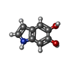  Mass: 149.147 Da / Num. of mol.: 2 / Source method: obtained synthetically / Formula: C8H7NO2 Mass: 149.147 Da / Num. of mol.: 2 / Source method: obtained synthetically / Formula: C8H7NO2#5: Water | ChemComp-HOH / |  Mass: 18.015 Da / Num. of mol.: 73 / Source method: isolated from a natural source / Formula: H2O Mass: 18.015 Da / Num. of mol.: 73 / Source method: isolated from a natural source / Formula: H2OHas protein modification | Y | |
-Experimental details
-Experiment
| Experiment | Method: X-RAY DIFFRACTION / Number of used crystals: 3 |
|---|
- Sample preparation
| Crystal | Density Matthews: 2.88 Å3/Da / Density % sol: 57.36 % | ||||||||||||||||||||
|---|---|---|---|---|---|---|---|---|---|---|---|---|---|---|---|---|---|---|---|---|---|
| Crystal grow | Temperature: 277 K / Method: microdialysis / pH: 5 Details: 20mM Tris-Hcl, pH 5.0 concentration 50 mg/ml 10% ethanol soaked for 12hours in buffer containing DOPA, MICRODIALYSIS, temperature 4K | ||||||||||||||||||||
| Crystal grow | *PLUS | ||||||||||||||||||||
| Components of the solutions | *PLUS
|
-Data collection
| Diffraction | Mean temperature: 283 K |
|---|---|
| Diffraction source | Source: ROTATING ANODE / Type: RIGAKU RU200 / Wavelength: 1.5418 |
| Detector | Type: MARRESEARCH / Detector: IMAGE PLATE / Date: May 7, 1998 |
| Radiation | Protocol: SINGLE WAVELENGTH / Monochromatic (M) / Laue (L): M / Scattering type: x-ray |
| Radiation wavelength | Wavelength: 1.5418 Å / Relative weight: 1 |
| Reflection | Resolution: 2.7→15 Å / Num. all: 18557 / Num. obs: 18557 / % possible obs: 91 % / Observed criterion σ(F): 0 / Observed criterion σ(I): 0 / Redundancy: 2.3 % / Biso Wilson estimate: 31 Å2 / Rmerge(I) obs: 0.124 / Net I/σ(I): 3.6 |
| Reflection shell | Resolution: 2.7→15 Å / Redundancy: 2.3 % / Rmerge(I) obs: 0.124 / Num. unique all: 18557 / % possible all: 91 |
| Reflection | *PLUS % possible obs: 91 % / Num. measured all: 100340 / Rmerge(I) obs: 0.071 |
| Reflection shell | *PLUS Lowest resolution: 2.9 Å / % possible obs: 63 % / Rmerge(I) obs: 0.142 |
- Processing
| Software |
| |||||||||||||||||||||||||
|---|---|---|---|---|---|---|---|---|---|---|---|---|---|---|---|---|---|---|---|---|---|---|---|---|---|---|
| Refinement | Resolution: 2.7→15 Å / σ(F): 0 / σ(I): 0 Stereochemistry target values: 84% most allowed region and 2 residues are in disallowed region 299, 640 due to gama turn.
| |||||||||||||||||||||||||
| Refinement step | Cycle: LAST / Resolution: 2.7→15 Å
| |||||||||||||||||||||||||
| Refine LS restraints |
| |||||||||||||||||||||||||
| Software | *PLUS Name: X-PLOR / Classification: refinement | |||||||||||||||||||||||||
| Refinement | *PLUS Highest resolution: 2.7 Å / Lowest resolution: 15 Å / σ(F): 0 / % reflection Rfree: 5 % | |||||||||||||||||||||||||
| Solvent computation | *PLUS | |||||||||||||||||||||||||
| Displacement parameters | *PLUS Biso mean: 31 Å2 | |||||||||||||||||||||||||
| Refine LS restraints | *PLUS
|
