 Movie
Movie Controller

[English] 日本語
 Yorodumi
- PDB-1jc1: CRYSTAL STRUCTURE ANALYSIS OF A REDOX-SENSITIVE GREEN FLUORESCENT... -
+ Open data

- Basic information
| Entry | Database: PDB / ID: 1jc1 | |||||||||
|---|---|---|---|---|---|---|---|---|---|---|
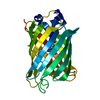
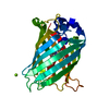
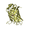
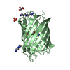
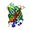
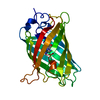
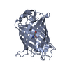
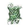
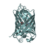
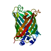
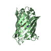
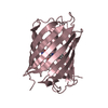
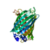
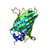
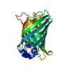
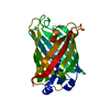
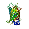
| Title | CRYSTAL STRUCTURE ANALYSIS OF A REDOX-SENSITIVE GREEN FLUORESCENT PROTEIN VARIANT IN A OXIDIZED FORM | |||||||||
 Components Components | GREEN FLUORESCENT PROTEIN | |||||||||
 Keywords Keywords | LUMINESCENT PROTEIN / Beta Barrel / Chromophore / Disulfide Bond | |||||||||
| Function / homology |  Function and homology information Function and homology information | |||||||||
| Biological species |   Aequorea victoria (jellyfish) Aequorea victoria (jellyfish) | |||||||||
| Method |  X-RAY DIFFRACTION / SYNCHROTRON / MOLECULAR REPLACEMENT / Resolution: 1.9 Å X-RAY DIFFRACTION / SYNCHROTRON / MOLECULAR REPLACEMENT / Resolution: 1.9 Å | |||||||||
 Authors Authors | Hanson, G.T. / Aggeler, R. / Oglesbee, D. / Cannon, M. / Capaldi, R.A. / Tsien, R.Y. / Remington, S.J. | |||||||||
 Citation Citation | Journal: J.Biol.Chem. / Year: 2004 Title: Investigating mitochondrial redox potential with redox-sensitive green fluorescent protein indicators. Authors: Hanson, G.T. / Aggeler, R. / Oglesbee, D. / Cannon, M. / Capaldi, R.A. / Tsien, R.Y. / Remington, S.J. | |||||||||
| History |
|
- Structure visualization
| Structure viewer | Molecule: MolmilJmol/JSmol |
|---|
- Downloads & links
-Download
| PDBx/mmCIF format | 1jc1.cif.gz | 148 KB | Display | PDBx/mmCIF format |
|---|---|---|---|---|
| PDB format | pdb1jc1.ent.gz | 115.3 KB | Display | PDB format |
| PDBx/mmJSON format | 1jc1.json.gz | Tree view | PDBx/mmJSON format | |
| Others |  Other downloads Other downloads |
-Validation report
| Arichive directory | https://data.pdbj.org/pub/pdb/validation_reports/jc/1jc1ftp://data.pdbj.org/pub/pdb/validation_reports/jc/1jc1 | HTTPS FTP |
|---|
-Related structure data
| Related structure data |  1jc0C  1emaS C: citing same article ( S: Starting model for refinement |
|---|---|
| Similar structure data |


-Links
 PDBj
PDBj

- Assembly
| Deposited unit | 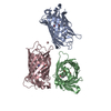
| ||||||||
|---|---|---|---|---|---|---|---|---|---|
| 1 | 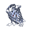
| ||||||||
| 2 | 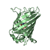
| ||||||||
| 3 | 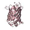
| ||||||||
| Unit cell |
|
-Components
| #1: Protein | Mass: 26886.379 Da / Num. of mol.: 3 / Mutation: C48S,F64L,S65T,Q80R,S147C,Q204C Source method: isolated from a genetically manipulated source Source: (gene. exp.) Aequorea victoria (jellyfish) / Plasmid: pRSETb / Production host:  #2: Water | ChemComp-HOH / |  Mass: 18.015 Da / Num. of mol.: 174 / Source method: isolated from a natural source / Formula: H2O Mass: 18.015 Da / Num. of mol.: 174 / Source method: isolated from a natural source / Formula: H2OHas protein modification | Y | |
|---|
-Experimental details
-Experiment
| Experiment | Method: X-RAY DIFFRACTION / Number of used crystals: 1 |
|---|
- Sample preparation
| Crystal | Density Matthews: 2.2 Å3/Da / Density % sol: 43.99 % | |||||||||||||||||||||||||||||||||||||||||||||||||
|---|---|---|---|---|---|---|---|---|---|---|---|---|---|---|---|---|---|---|---|---|---|---|---|---|---|---|---|---|---|---|---|---|---|---|---|---|---|---|---|---|---|---|---|---|---|---|---|---|---|---|
| Crystal grow | Temperature: 295 K / Method: vapor diffusion, hanging drop / pH: 8.5 Details: PEG 4000, Lithium Sulfate, Tris, Copper (II) Chloride, pH 8.5, VAPOR DIFFUSION, HANGING DROP, temperature 295K | |||||||||||||||||||||||||||||||||||||||||||||||||
| Crystal grow | *PLUS pH: 7.9 / Method: vapor diffusion, hanging drop | |||||||||||||||||||||||||||||||||||||||||||||||||
| Components of the solutions | *PLUS
|
-Data collection
| Diffraction | Mean temperature: 100 K |
|---|---|
| Diffraction source | Source: SYNCHROTRON / Site: SSRL  / Beamline: BL7-1 / Wavelength: 1.08 Å / Beamline: BL7-1 / Wavelength: 1.08 Å |
| Detector | Type: MAR scanner 345 mm plate / Detector: IMAGE PLATE / Date: Nov 25, 2000 Details: 58 cm long, Pt-coated fused silica, vertical focus mirror |
| Radiation | Monochromator: Cylindrically bent triangular Si(111) asymmetric cut horizontal focus Protocol: SINGLE WAVELENGTH / Monochromatic (M) / Laue (L): M / Scattering type: x-ray |
| Radiation wavelength | Wavelength: 1.08 Å / Relative weight: 1 |
| Reflection | Resolution: 1.9→29.7 Å / Num. all: 203865 / Num. obs: 56854 / % possible obs: 99.8 % / Redundancy: 3.6 % / Biso Wilson estimate: 27.7 Å2 / Rmerge(I) obs: 0.058 / Net I/σ(I): 7.2 |
| Reflection shell | Resolution: 1.9→1.94 Å / Redundancy: 3.6 % / Rmerge(I) obs: 0.314 / Mean I/σ(I) obs: 2.3 / Num. unique all: 4181 / % possible all: 100 |
| Reflection | *PLUS Num. measured all: 203865 |
| Reflection shell | *PLUS Lowest resolution: 1.95 Å / % possible obs: 100 % / Rmerge(I) obs: 0.247 |
- Processing
| Software |
| ||||||||||||||||||
|---|---|---|---|---|---|---|---|---|---|---|---|---|---|---|---|---|---|---|---|
| Refinement | Method to determine structure: MOLECULAR REPLACEMENT Starting model: PDB ENTRY 1EMA Resolution: 1.9→30 Å / Stereochemistry target values: ENGH & HUBER /
| ||||||||||||||||||
| Refinement step | Cycle: LAST / Resolution: 1.9→30 Å
| ||||||||||||||||||
| Refine LS restraints |
| ||||||||||||||||||
| LS refinement shell | Resolution: 1.9→1.94 Å
| ||||||||||||||||||
| Refinement | *PLUS Highest resolution: 1.9 Å / Lowest resolution: 6 Å / Num. reflection obs: 54921 / Num. reflection Rfree: 5471 / Rfactor Rfree: 0.226 / Rfactor Rwork: 0.317 | ||||||||||||||||||
| Solvent computation | *PLUS | ||||||||||||||||||
| Displacement parameters | *PLUS | ||||||||||||||||||
| Refine LS restraints | *PLUS
|
