 Movie
Movie Controller


+ Open data

- Basic information
| Entry | Database: PDB / ID: 1ema | |||||||||
|---|---|---|---|---|---|---|---|---|---|---|
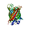
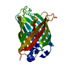
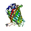
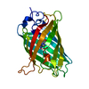
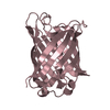
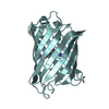
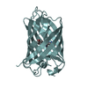
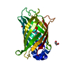
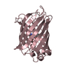
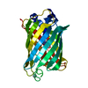
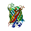
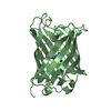
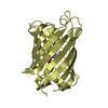
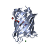
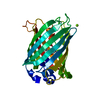
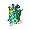
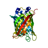
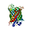
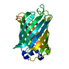
| Title | GREEN FLUORESCENT PROTEIN FROM AEQUOREA VICTORIA | |||||||||
 Components Components | GREEN FLUORESCENT PROTEIN | |||||||||
 Keywords Keywords | FLUORESCENT PROTEIN / BETA-BARREL / AUTOCATALYTIC / FLUOROPHORE / BIOLUMINESCENSE FLUORESCENT PROTEIN | |||||||||
| Function / homology |  Function and homology information Function and homology information | |||||||||
| Biological species |   Aequorea victoria (jellyfish) Aequorea victoria (jellyfish) | |||||||||
| Method |  X-RAY DIFFRACTION / SYNCHROTRON / MIR/SAD / Resolution: 1.9 Å X-RAY DIFFRACTION / SYNCHROTRON / MIR/SAD / Resolution: 1.9 Å | |||||||||
 Authors Authors | Ormo, M. / Remington, S.J. | |||||||||
 Citation Citation | Journal: Science / Year: 1996 Title: Crystal structure of the Aequorea victoria green fluorescent protein. Authors: Ormo, M. / Cubitt, A.B. / Kallio, K. / Gross, L.A. / Tsien, R.Y. / Remington, S.J. #1: Journal: Nature / Year: 1995Title: Improved Green Fluorescence Authors: Heim, R. / Cubitt, A.B. / Tsien, R.Y. | |||||||||
| History |
|
- Structure visualization
| Structure viewer | Molecule: MolmilJmol/JSmol |
|---|
- Downloads & links
-Download
| PDBx/mmCIF format | 1ema.cif.gz | 60.1 KB | Display | PDBx/mmCIF format |
|---|---|---|---|---|
| PDB format | pdb1ema.ent.gz | 43.1 KB | Display | PDB format |
| PDBx/mmJSON format | 1ema.json.gz | Tree view | PDBx/mmJSON format | |
| Others |  Other downloads Other downloads |
-Validation report
| Arichive directory | https://data.pdbj.org/pub/pdb/validation_reports/em/1emaftp://data.pdbj.org/pub/pdb/validation_reports/em/1ema | HTTPS FTP |
|---|
-Related structure data
| Similar structure data |
|---|

-Links
 PDBj
PDBj

- Assembly
| Deposited unit | 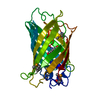
| ||||||||
|---|---|---|---|---|---|---|---|---|---|
| 1 |
| ||||||||
| Unit cell |
|
-Components
| #1: Protein | Mass: 27226.754 Da / Num. of mol.: 1 / Mutation: 65 - 67 REPLACED BY CRO, Q80R Source method: isolated from a genetically manipulated source Details: THE PROTEIN CONTAINS SIX SE-METHIONINES. OF THESE, THE N-TERMINAL MET AND MET 233 ARE NOT PRESENT IN THE ENTRY Source: (gene. exp.) Aequorea victoria (jellyfish) / Description: THE N-TERMINAL HIS-TAG HAS BEEN REMOVED / Organ: LEAVES / Plasmid: PRSETB (INVITROGEN) / Production host:  |
|---|---|
| #2: Water | ChemComp-HOH /  Mass: 18.015 Da / Num. of mol.: 95 / Source method: isolated from a natural source / Formula: H2O Mass: 18.015 Da / Num. of mol.: 95 / Source method: isolated from a natural source / Formula: H2O |
| Has protein modification | Y |
| Sequence details | THE FLUOROPHORE (CRO) IS GENERATED BY AN AUTOCATALYTIC CYCLIZATION OF THE POLYPEPTIDE BACKBONE ...THE FLUOROPHOR |
-Experimental details
-Experiment
| Experiment | Method: X-RAY DIFFRACTION / Number of used crystals: 1 |
|---|
- Sample preparation
| Crystal | Density Matthews: 2.1 Å3/Da / Density % sol: 40.5 % | ||||||||||||||||||||||||||||||
|---|---|---|---|---|---|---|---|---|---|---|---|---|---|---|---|---|---|---|---|---|---|---|---|---|---|---|---|---|---|---|---|
| Crystal grow | pH: 8.2 Details: 22-26% PEG 4000, 50 MM HEPES PH 8.0-8.4, 50 MM MGCL2, 10 MM 2-MERCAPTOETHANOL, 5-7 MG PROTEIN, pH 8.2 PH range: 8.0-8.4 | ||||||||||||||||||||||||||||||
| Crystal grow | *PLUS Method: vapor diffusion, hanging drop | ||||||||||||||||||||||||||||||
| Components of the solutions | *PLUS
|
-Data collection
| Diffraction | Mean temperature: 295 K |
|---|---|
| Diffraction source | Source: SYNCHROTRON / Site: NSLS  / Beamline: X4A / Wavelength: 0.979 / Beamline: X4A / Wavelength: 0.979 |
| Detector | Type: FUJI / Detector: IMAGE PLATE / Date: Apr 28, 1996 |
| Radiation | Monochromatic (M) / Laue (L): M / Scattering type: x-ray |
| Radiation wavelength | Wavelength: 0.979 Å / Relative weight: 1 |
| Reflection | Highest resolution: 1.9 Å / Num. obs: 17676 / % possible obs: 84 % / Observed criterion σ(I): 0 / Redundancy: 6.45 % / Biso Wilson estimate: 16.5 Å2 / Rmerge(I) obs: 0.09 |
- Processing
| Software |
| ||||||||||||||||||||||||||||||||||||||||||||||||||
|---|---|---|---|---|---|---|---|---|---|---|---|---|---|---|---|---|---|---|---|---|---|---|---|---|---|---|---|---|---|---|---|---|---|---|---|---|---|---|---|---|---|---|---|---|---|---|---|---|---|---|---|
| Refinement | Method to determine structure: MIR/SAD / Resolution: 1.9→20 Å / Isotropic thermal model: TNT / σ(F): 0 / Stereochemistry target values: TNT Details: THE FINAL (FO-FC) DENSITY SHOWS LARGE DIFFERENCE FEATURES LOCATED AROUND THE MAIN CHAIN PART OF THE FLUOROPHORE THAT ORIGINATES FROM RESIDUES THR 65 AND GLY 67. THE DIFFERENCE DENSITIES ALSO ...Details: THE FINAL (FO-FC) DENSITY SHOWS LARGE DIFFERENCE FEATURES LOCATED AROUND THE MAIN CHAIN PART OF THE FLUOROPHORE THAT ORIGINATES FROM RESIDUES THR 65 AND GLY 67. THE DIFFERENCE DENSITIES ALSO AFFECT THE ATOMIC-POSITION REFINEMENT OF VAL 68. THE DIFFERENCE FEATURES MIGHT BE EXPLAINED AS A SUBSET (<30%) OF MOLECULES THAT HAS FAILED TO UNDERGO THE COMPLETE FORMATION OF THE FLUOROPHORE. THE LOOP RESIDUES 157 AND 158 ARE DISORDERED. A NUMBER OF SURFACE RESIDUES HAVE TRUNCATED SIDE CHAINS DUE TO WEAK OR NO DENSITY.
| ||||||||||||||||||||||||||||||||||||||||||||||||||
| Solvent computation | Solvent model: BABINET SCALING / Bsol: 300 Å2 / ksol: 0.8 e/Å3 | ||||||||||||||||||||||||||||||||||||||||||||||||||
| Refinement step | Cycle: LAST / Resolution: 1.9→20 Å
| ||||||||||||||||||||||||||||||||||||||||||||||||||
| Refine LS restraints |
| ||||||||||||||||||||||||||||||||||||||||||||||||||
| Software | *PLUS Name: TNT / Version: 5-F / Classification: refinement | ||||||||||||||||||||||||||||||||||||||||||||||||||
| Refinement | *PLUS Rfactor obs: 0.175 | ||||||||||||||||||||||||||||||||||||||||||||||||||
| Solvent computation | *PLUS | ||||||||||||||||||||||||||||||||||||||||||||||||||
| Displacement parameters | *PLUS Biso mean: 24.1 Å2 | ||||||||||||||||||||||||||||||||||||||||||||||||||
| Refine LS restraints | *PLUS
|
