 Movie
Movie Controller

[English] 日本語
 Yorodumi
- PDB-2hgy: Structure of S65A Y66F E222A GFP variant after cyclization and ca... -
+ Open data

- Basic information
| Entry | Database: PDB / ID: 2hgy | |||||||||
|---|---|---|---|---|---|---|---|---|---|---|
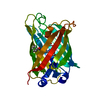
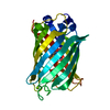
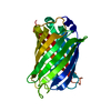
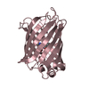
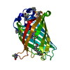
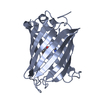
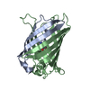
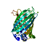
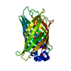
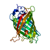
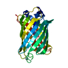
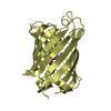
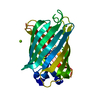
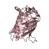
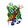
| Title | Structure of S65A Y66F E222A GFP variant after cyclization and carbon-carbon bond cleavage | |||||||||
 Components Components | Green fluorescent protein | |||||||||
 Keywords Keywords | LUMINESCENT PROTEIN / post-translational modification / cyclization / carbon-carbon bond cleavage / radical / GFP | |||||||||
| Function / homology |  Function and homology information Function and homology information | |||||||||
| Biological species |   Aequorea victoria (jellyfish) Aequorea victoria (jellyfish) | |||||||||
| Method |  X-RAY DIFFRACTION / SYNCHROTRON / MOLECULAR REPLACEMENT / Resolution: 2.05 Å X-RAY DIFFRACTION / SYNCHROTRON / MOLECULAR REPLACEMENT / Resolution: 2.05 Å | |||||||||
 Authors Authors | Barondeau, D.P. / Kassmann, C.J. / Tainer, J.A. / Getzoff, E.D. | |||||||||
 Citation Citation | Journal: J.Am.Chem.Soc. / Year: 2007 Title: The Case of the Missing Ring: Radical Cleavage of a Carbon-Carbon Bond and Implications for GFP Chromophore Biosynthesis Authors: Barondeau, D.P. / Kassmann, C.J. / Tainer, J.A. / Getzoff, E.D. | |||||||||
| History |
|
- Structure visualization
| Structure viewer | Molecule: MolmilJmol/JSmol |
|---|
- Downloads & links
-Download
| PDBx/mmCIF format | 2hgy.cif.gz | 60.7 KB | Display | PDBx/mmCIF format |
|---|---|---|---|---|
| PDB format | pdb2hgy.ent.gz | 43.4 KB | Display | PDB format |
| PDBx/mmJSON format | 2hgy.json.gz | Tree view | PDBx/mmJSON format | |
| Others |  Other downloads Other downloads |
-Validation report
| Arichive directory | https://data.pdbj.org/pub/pdb/validation_reports/hg/2hgyftp://data.pdbj.org/pub/pdb/validation_reports/hg/2hgy | HTTPS FTP |
|---|
-Related structure data
| Related structure data |  2hcgC  2hfcC  2hgdC  1emaS C: citing same article ( S: Starting model for refinement |
|---|---|
| Similar structure data |


-Links
 PDBj
PDBj

- Assembly
| Deposited unit | 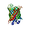
| ||||||||
|---|---|---|---|---|---|---|---|---|---|
| 1 |
| ||||||||
| Unit cell |
|
-Components
| #1: Protein | Mass: 26676.986 Da / Num. of mol.: 1 / Mutation: S65A, Y66F, F99S, M153T, V163A, E222A Source method: isolated from a genetically manipulated source Source: (gene. exp.) Aequorea victoria (jellyfish) / Gene: GFP / Plasmid: pET11a / Production host:  |
|---|---|
| #2: Water | ChemComp-HOH /  Mass: 18.015 Da / Num. of mol.: 130 / Source method: isolated from a natural source / Formula: H2O Mass: 18.015 Da / Num. of mol.: 130 / Source method: isolated from a natural source / Formula: H2O |
| Has protein modification | Y |
| Sequence details | RESIDUE AT POSITION 65 IS A S65A MUTATION, RESIDUE AT POSITION 66 IS A Y66F MUTATION. RESIDUES ALA ...RESIDUE AT POSITION 65 IS A S65A MUTATION, RESIDUE AT POSITION 66 IS A Y66F MUTATION. RESIDUES ALA 65, PHE 66 AND GLY 67 CONSTITUTE |
-Experimental details
-Experiment
| Experiment | Method: X-RAY DIFFRACTION / Number of used crystals: 1 |
|---|
- Sample preparation
| Crystal | Density Matthews: 2.08 Å3/Da / Density % sol: 40.77 % |
|---|---|
| Crystal grow | Temperature: 298 K / Method: vapor diffusion, hanging drop / pH: 8 Details: 19% PEG 4K, 50 mM MgCl2, 50 mM Hepes, pH 8.0, VAPOR DIFFUSION, HANGING DROP, temperature 298K |
-Data collection
| Diffraction | Mean temperature: 100 K |
|---|---|
| Diffraction source | Source: SYNCHROTRON / Site: SSRL  / Beamline: BL11-1 / Wavelength: 0.979 Å / Beamline: BL11-1 / Wavelength: 0.979 Å |
| Detector | Type: ADSC QUANTUM 315 / Detector: CCD / Date: Mar 10, 2003 |
| Radiation | Protocol: SINGLE WAVELENGTH / Monochromatic (M) / Laue (L): M / Scattering type: x-ray |
| Radiation wavelength | Wavelength: 0.979 Å / Relative weight: 1 |
| Reflection | Resolution: 2.05→20 Å / Num. obs: 13519 / % possible obs: 93.1 % / Observed criterion σ(F): 0 / Observed criterion σ(I): -3 / Biso Wilson estimate: 25.2 Å2 / Rsym value: 0.07 / Net I/σ(I): 20.2 |
| Reflection shell | Resolution: 2.05→2.1 Å / Mean I/σ(I) obs: 4.8 / Num. unique all: 1204 / Rsym value: 0.357 / % possible all: 85.2 |
- Processing
| Software |
| |||||||||||||||||||||||||
|---|---|---|---|---|---|---|---|---|---|---|---|---|---|---|---|---|---|---|---|---|---|---|---|---|---|---|
| Refinement | Method to determine structure: MOLECULAR REPLACEMENT Starting model: PDB ENTRY 1ema Resolution: 2.05→20 Å / Cross valid method: THROUGHOUT / σ(F): 0 / σ(I): 0 / Stereochemistry target values: Engh & Huber
| |||||||||||||||||||||||||
| Refinement step | Cycle: LAST / Resolution: 2.05→20 Å
| |||||||||||||||||||||||||
| Refine LS restraints |
|
