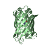
 Movie
Movie Controller


+ Open data

- Basic information
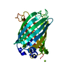
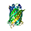
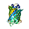
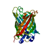
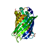
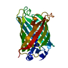
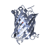
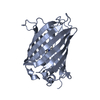
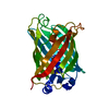
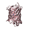
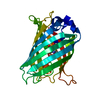
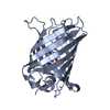
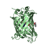
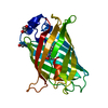
| Entry | Database: PDB / ID: 2g16 | |||||||||
|---|---|---|---|---|---|---|---|---|---|---|
| Title | Structure of S65A Y66S GFP variant after backbone fragmentation | |||||||||
 Components Components | (Green fluorescent protein) x 2 | |||||||||
 Keywords Keywords | LUMINESCENT PROTEIN / Beta barrel / chromophore / biosynthesis / fragmentaion / denaturation | |||||||||
| Function / homology |  Function and homology information Function and homology information | |||||||||
| Biological species |   Aequorea victoria (jellyfish) Aequorea victoria (jellyfish) | |||||||||
| Method |  X-RAY DIFFRACTION / SYNCHROTRON / MOLECULAR REPLACEMENT / Resolution: 2 Å X-RAY DIFFRACTION / SYNCHROTRON / MOLECULAR REPLACEMENT / Resolution: 2 Å | |||||||||
 Authors Authors | Barondeau, D.P. | |||||||||
 Citation Citation | Journal: J.Am.Chem.Soc. / Year: 2006 Title: Understanding GFP Posttranslational Chemistry: Structures of Designed Variants that Achieve Backbone Fragmentation, Hydrolysis, and Decarboxylation. Authors: Barondeau, D.P. / Kassmann, C.J. / Tainer, J.A. / Getzoff, E.D. | |||||||||
| History |
| |||||||||
| Remark 999 | SEQUENCE Ser 65 in the database has been mutated to alanine, Tyr 66 in the database has been ...SEQUENCE Ser 65 in the database has been mutated to alanine, Tyr 66 in the database has been mutated to serine. Residues Ala 65, Ser 66 and Gly 67 consitute the chromophore CRW 66. This ligand underwent denaturation-induced backbone fragmentation between the C1 and CA1 atoms resulting in fragmented CRW ligand and an E1H molecule |
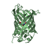
- Structure visualization
| Structure viewer | Molecule: MolmilJmol/JSmol |
|---|
- Downloads & links
-Download
| PDBx/mmCIF format | 2g16.cif.gz | 63 KB | Display | PDBx/mmCIF format |
|---|---|---|---|---|
| PDB format | pdb2g16.ent.gz | 44.1 KB | Display | PDB format |
| PDBx/mmJSON format | 2g16.json.gz | Tree view | PDBx/mmJSON format | |
| Others |  Other downloads Other downloads |
-Validation report
| Arichive directory | https://data.pdbj.org/pub/pdb/validation_reports/g1/2g16ftp://data.pdbj.org/pub/pdb/validation_reports/g1/2g16 | HTTPS FTP |
|---|
-Related structure data
| Related structure data |  2g2sC 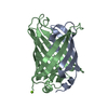 2g3dC  2g5zC  2g6eC  1emaS S: Starting model for refinement C: citing same article ( |
|---|---|
| Similar structure data |


-Links
 PDBj
PDBj

- Assembly
| Deposited unit | 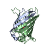
| ||||||||
|---|---|---|---|---|---|---|---|---|---|
| 1 |
| ||||||||
| Unit cell |
|
-Components
| #1: Protein | Mass: 6930.946 Da / Num. of mol.: 1 / Fragment: residues 2-64 Source method: isolated from a genetically manipulated source Source: (gene. exp.) Aequorea victoria (jellyfish) / Gene: GFP / Plasmid: pET11a / Species (production host): Escherichia coli / Production host:  |
|---|---|
| #2: Protein | Mass: 19818.086 Da / Num. of mol.: 1 / Fragment: residues 65-238 / Mutation: S65A, Y66S, F99S, M153T, V163A Source method: isolated from a genetically manipulated source Source: (gene. exp.) Aequorea victoria (jellyfish) / Gene: GFP / Plasmid: pET11a / Species (production host): Escherichia coli / Production host: |
| #3: Water | ChemComp-HOH /  Mass: 18.015 Da / Num. of mol.: 173 / Source method: isolated from a natural source / Formula: H2O Mass: 18.015 Da / Num. of mol.: 173 / Source method: isolated from a natural source / Formula: H2O |
-Experimental details
-Experiment
| Experiment | Method: X-RAY DIFFRACTION / Number of used crystals: 1 |
|---|
- Sample preparation
| Crystal | Density Matthews: 2.1 Å3/Da / Density % sol: 41.3 % |
|---|---|
| Crystal grow | Temperature: 298 K / Method: vapor diffusion, hanging drop / pH: 8 Details: 50 mM MgCl2, 50 mM Hepes, 20% PEG 4000, pH 8.0, VAPOR DIFFUSION, HANGING DROP, temperature 298K |
-Data collection
| Diffraction | Mean temperature: 100 K |
|---|---|
| Diffraction source | Source: SYNCHROTRON / Site: SSRL  / Beamline: BL11-1 / Wavelength: 0.85 Å / Beamline: BL11-1 / Wavelength: 0.85 Å |
| Detector | Type: ADSC QUANTUM 315 / Detector: CCD / Date: Mar 3, 2002 |
| Radiation | Protocol: SINGLE WAVELENGTH / Monochromatic (M) / Laue (L): M / Scattering type: x-ray |
| Radiation wavelength | Wavelength: 0.85 Å / Relative weight: 1 |
| Reflection | Resolution: 2→20 Å / Num. all: 15928 / Num. obs: 15582 / % possible obs: 97.8 % / Observed criterion σ(I): -3 / Biso Wilson estimate: 19 Å2 / Rsym value: 0.075 / Net I/σ(I): 16 |
| Reflection shell | Resolution: 2→2.07 Å / Mean I/σ(I) obs: 3.9 / Rsym value: 0.254 / % possible all: 97.1 |
- Processing
| Software |
| ||||||||||||||||||||
|---|---|---|---|---|---|---|---|---|---|---|---|---|---|---|---|---|---|---|---|---|---|
| Refinement | Method to determine structure: MOLECULAR REPLACEMENT Starting model: PDB ENTRY 1ema Resolution: 2→20 Å / Cross valid method: THROUGHOUT / σ(F): 0 / Stereochemistry target values: Engh & Huber
| ||||||||||||||||||||
| Refinement step | Cycle: LAST / Resolution: 2→20 Å
| ||||||||||||||||||||
| Refine LS restraints |
| ||||||||||||||||||||
| LS refinement shell | Resolution: 2→2.03 Å /
|
