[English] 日本語
Yorodumi
- PDB-6vio: Crystal structure of eYFP His148Ser -

+
Open data


ID or keywords:

Loading...

-
Basic information

Entry
Database: PDB / ID: 6vio
TitleCrystal structure of eYFP His148Ser
ComponentsGreen fluorescent protein GFP
Keywords
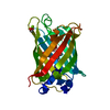
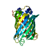
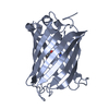
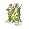
FLUORESCENT PROTEIN / yellow / enhanced / modulatable
Function / homology
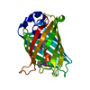
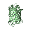
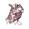
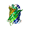
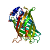
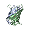
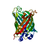
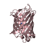
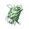
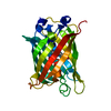
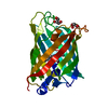
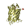
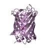
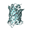
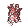
Green fluorescent protein, GFP / Green fluorescent protein-related / Green fluorescent protein / Green fluorescent protein / bioluminescence / generation of precursor metabolites and energy / Green fluorescent protein GFP / Green fluorescent protein
Function and homology information
Biological speciesAequorea victoria (jellyfish)
MethodX-RAY DIFFRACTION / SYNCHROTRON / MOLECULAR REPLACEMENT / molecular replacement / Resolution: 3.6 Å
AuthorsLieberman, R.L. / Hill, S.E. / Patterson-Orazem, A.C.
Funding support United States, 1items
OrganizationGrant numberCountry
National Institutes of Health/National Eye Institute (NIH/NEI)R01EY021205 United States
CitationJournal: J.Phys.Chem.B / Year: 2021
Title: Optically Modulated and Optically Activated Delayed Fluorescent Proteins through Dark State Engineering
Authors: Peng, B. / Dikdan, R. / Hill, S.E. / Patterson-Orazem, A.C. / Lieberman, R.L. / Fahrni, C.J. / Dickson, R.M.
History
DepositionJan 13, 2020Deposition site: RCSB / Processing site: RCSB
Revision 1.0Jan 13, 2021Provider: repository / Type: Initial release
Revision 1.1Jul 28, 2021Group: Database references / Category: citation / citation_author
Item: _citation.country / _citation.journal_abbrev ..._citation.country / _citation.journal_abbrev / _citation.journal_id_ASTM / _citation.journal_id_CSD / _citation.journal_id_ISSN / _citation.journal_volume / _citation.page_first / _citation.page_last / _citation.pdbx_database_id_DOI / _citation.pdbx_database_id_PubMed / _citation.title / _citation.year
Revision 1.2Oct 11, 2023Group: Data collection / Database references / Refinement description
Category: chem_comp_atom / chem_comp_bond ...chem_comp_atom / chem_comp_bond / database_2 / pdbx_initial_refinement_model / struct_ncs_dom_lim
Item: _database_2.pdbx_DOI / _database_2.pdbx_database_accession ..._database_2.pdbx_DOI / _database_2.pdbx_database_accession / _struct_ncs_dom_lim.beg_auth_comp_id / _struct_ncs_dom_lim.beg_label_asym_id / _struct_ncs_dom_lim.beg_label_comp_id / _struct_ncs_dom_lim.beg_label_seq_id / _struct_ncs_dom_lim.end_auth_comp_id / _struct_ncs_dom_lim.end_label_asym_id / _struct_ncs_dom_lim.end_label_comp_id / _struct_ncs_dom_lim.end_label_seq_id
Revision 1.3Nov 15, 2023Group: Data collection / Category: chem_comp_atom / chem_comp_bond / Item: _chem_comp_atom.atom_id / _chem_comp_bond.atom_id_2

-
Structure visualization

Structure viewerMolecule:
MolmilJmol/JSmol

Downloads & links

-
Assembly

Deposited unit
A: Green fluorescent protein GFP
B: Green fluorescent protein GFP
C: Green fluorescent protein GFP
D: Green fluorescent protein GFP
E: Green fluorescent protein GFP
F: Green fluorescent protein GFP
G: Green fluorescent protein GFP
H: Green fluorescent protein GFP


Theoretical massNumber of molelcules
Total (without water)235,3298
Polymers235,3298
Non-polymers00
Water00
1
A: Green fluorescent protein GFP


Theoretical massNumber of molelcules
Total (without water)29,4161
Polymers29,4161
Non-polymers00
Water0
TypeNameSymmetry operationNumber
identity operation1_555x,y,z1
MethodPISA
2
B: Green fluorescent protein GFP


Theoretical massNumber of molelcules
Total (without water)29,4161
Polymers29,4161
Non-polymers00
Water0
TypeNameSymmetry operationNumber
identity operation1_555x,y,z1
MethodPISA
3
C: Green fluorescent protein GFP


Theoretical massNumber of molelcules
Total (without water)29,4161
Polymers29,4161
Non-polymers00
Water0
TypeNameSymmetry operationNumber
identity operation1_555x,y,z1
MethodPISA
4
D: Green fluorescent protein GFP


Theoretical massNumber of molelcules
Total (without water)29,4161
Polymers29,4161
Non-polymers00
Water0
TypeNameSymmetry operationNumber
identity operation1_555x,y,z1
MethodPISA
5
E: Green fluorescent protein GFP


Theoretical massNumber of molelcules
Total (without water)29,4161
Polymers29,4161
Non-polymers00
Water0
TypeNameSymmetry operationNumber
identity operation1_555x,y,z1
MethodPISA
6
F: Green fluorescent protein GFP


Theoretical massNumber of molelcules
Total (without water)29,4161
Polymers29,4161
Non-polymers00
Water0
TypeNameSymmetry operationNumber
identity operation1_555x,y,z1
MethodPISA
7
G: Green fluorescent protein GFP


Theoretical massNumber of molelcules
Total (without water)29,4161
Polymers29,4161
Non-polymers00
Water0
TypeNameSymmetry operationNumber
identity operation1_555x,y,z1
MethodPISA
8
H: Green fluorescent protein GFP


Theoretical massNumber of molelcules
Total (without water)29,4161
Polymers29,4161
Non-polymers00
Water0
TypeNameSymmetry operationNumber
identity operation1_555x,y,z1
MethodPISA
Unit cell
Length a, b, c (Å)56.651, 56.772, 148.193
Angle α, β, γ (deg.)85.120, 85.260, 70.480
Int Tables number1
Space group name H-MP1
Noncrystallographic symmetry (NCS)NCS domain:
IDEns-IDDetails
11(chain A and resid 6 through 230)
21(chain B and resid 6 through 230)
31(chain C and resid 6 through 230)
41(chain D and resid 6 through 230)
51(chain E and resid 6 through 230)
61(chain F and resid 6 through 230)
71(chain G and resid 6 through 230)
81(chain H and resid 6 through 230)

NCS domain segments:

Component-ID: 1 / Ens-ID: 1 / Beg auth comp-ID: GLU / Beg label comp-ID: GLU / End auth comp-ID: THR / End label comp-ID: THR / Auth seq-ID: 6 - 230 / Label seq-ID: 30 - 252

Dom-IDSelection detailsAuth asym-IDLabel asym-ID
1(chain A and resid 6 through 230)AA
2(chain B and resid 6 through 230)BB
3(chain C and resid 6 through 230)CC
4(chain D and resid 6 through 230)DD
5(chain E and resid 6 through 230)EE
6(chain F and resid 6 through 230)FF
7(chain G and resid 6 through 230)GG
8(chain H and resid 6 through 230)HH

-
Components

#1: Protein
Green fluorescent protein GFP


Mass: 29416.145 Da / Num. of mol.: 8
Source method: isolated from a genetically manipulated source
Source: (gene. exp.) Aequorea victoria (jellyfish) / Production host: Escherichia coli (E. coli) / References: UniProt: A0A5J6CYR6, UniProt: P42212*PLUS
Has ligand of interestY

-
Experimental details

-
Experiment

ExperimentMethod: X-RAY DIFFRACTION / Number of used crystals: 1

-
Sample preparation

CrystalDescription: plate
Crystal growTemperature: 298 K / Method: vapor diffusion, hanging drop / Details: 1.75 M Na/K phosphate pH 6.9

-
Data collection

DiffractionMean temperature: 100 K / Serial crystal experiment: N
Diffraction sourceSource: SYNCHROTRON / Site: APS / Beamline: 22-ID / Wavelength: 1 Å
DetectorType: DECTRIS EIGER X 16M / Detector: PIXEL / Date: Apr 20, 2019
RadiationProtocol: SINGLE WAVELENGTH / Monochromatic (M) / Laue (L): M / Scattering type: x-ray
Radiation wavelengthWavelength: 1 Å / Relative weight: 1
ReflectionResolution: 3.6→50 Å / Num. obs: 18449 / % possible obs: 91.4 % / Redundancy: 2.2 % / Rmerge(I) obs: 0.161 / Rpim(I) all: 0.128 / Rrim(I) all: 0.207 / Χ2: 1.329 / Net I/σ(I): 4.4 / Num. measured all: 40830
Reflection shell

Diffraction-ID: 1

Resolution (Å)Redundancy (%)Rmerge(I) obsNum. unique obsCC1/2Rpim(I) allRrim(I) allΧ2% possible all
3.6-3.662.40.8539440.7630.6221.0620.60997.9
3.66-3.732.40.7510040.7410.5560.9390.77796.4
3.73-3.82.40.6710390.7860.4910.8360.73397.8
3.8-3.882.30.5239220.840.3930.6580.76496.4
3.88-3.962.30.56810060.8230.4260.7140.89496.5
3.96-4.052.40.3779560.9060.2810.4730.9496.7
4.05-4.162.30.3429370.9160.2590.4321.23394.8
4.16-4.272.30.2929710.9410.230.3741.21995.1
4.27-4.392.20.2529760.9450.1940.321.23293.1
4.39-4.542.20.2128880.960.1740.2761.52591.7
4.54-4.72.10.1849040.9640.150.2381.27789.9
4.7-4.892.10.1729250.9650.140.2231.39490.7
4.89-5.112.10.1488820.9780.1210.1921.45687.2
5.11-5.3820.1378680.9850.1140.1791.51287
5.38-5.711.90.1479000.9780.1220.1931.49186.5
5.71-6.1520.1548700.9680.1240.1991.6986.6
6.15-6.772.30.1168960.9920.0890.1471.65290.5
6.77-7.752.30.0878920.9910.070.1131.7788.8
7.75-9.752.20.0718530.990.0620.0952.72383.5
9.75-502.10.0568160.9920.050.0753.05881

-
Phasing

PhasingMethod: molecular replacement
Phasing MR
Highest resolutionLowest resolution
Rotation3.51 Å41.01 Å
Translation3.51 Å41.01 Å

-
Processing

Software
NameVersionClassification
HKL-2000data reduction
SCALEPACKdata scaling
PHASER2.8.2phasing
PHENIX1.15.2refinement
PDB_EXTRACT3.25data extraction
RefinementMethod to determine structure: MOLECULAR REPLACEMENT
Starting model: 3V3D
Resolution: 3.6→37.38 Å / SU ML: 0.53 / Cross valid method: THROUGHOUT / σ(F): 1.98 / Phase error: 40.23
RfactorNum. reflection% reflection
Rfree0.3101 1744 9.91 %
Rwork0.2372 --
obs0.245 17595 87.82 %
Solvent computationShrinkage radii: 0.9 Å / VDW probe radii: 1.11 Å
Displacement parametersBiso max: 178.93 Å2 / Biso mean: 104.1115 Å2 / Biso min: 66.38 Å2
Refinement stepCycle: final / Resolution: 3.6→37.38 Å
ProteinNucleic acidLigandSolventTotal
Num. atoms14010 0 0 0 14010
Num. residues----1809
Refine LS restraints NCS
Ens-IDDom-IDAuth asym-IDNumberRefine-IDRmsType
11A5360X-RAY DIFFRACTION0.921TORSIONAL
12B5360X-RAY DIFFRACTION0.921TORSIONAL
13C5360X-RAY DIFFRACTION0.921TORSIONAL
14D5360X-RAY DIFFRACTION0.921TORSIONAL
15E5360X-RAY DIFFRACTION0.921TORSIONAL
16F5360X-RAY DIFFRACTION0.921TORSIONAL
17G5360X-RAY DIFFRACTION0.921TORSIONAL
18H5360X-RAY DIFFRACTION0.921TORSIONAL
LS refinement shell

Refine-ID: X-RAY DIFFRACTION / Rfactor Rfree error: 0

Resolution (Å)Rfactor RfreeNum. reflection RfreeRfactor RworkNum. reflection Rwork% reflection obs (%)
3.6001-3.7060.34091350.342122982
3.706-3.82550.3931660.3252145796
3.8255-3.96210.34451520.2928143096
3.9621-4.12050.28551570.248142895
4.1205-4.30780.35071580.2533139993
4.3078-4.53460.26651490.2371134090
4.5346-4.81810.28781480.222135289
4.8181-5.18920.29271360.2204127684
5.1892-5.70980.27641400.2324125982
5.7098-6.53220.34181340.2535125185
6.5322-8.21550.29921430.238134189
8.2155-37.380.3081260.1689108973

+
About Yorodumi

-
News

-
Feb 9, 2022. New format data for meta-information of EMDB entries

New format data for meta-information of EMDB entries

  • Version 3 of the EMDB header file is now the official format.
  • The previous official version 1.9 will be removed from the archive.

Related info.:EMDB header

External links:wwPDB to switch to version 3 of the EMDB data model

-
Aug 12, 2020. Covid-19 info

Covid-19 info

URL: https://pdbjlvh1.pdbj.org/emnavi/covid19.php

New page: Covid-19 featured information page in EM Navigator.

Related info.:Covid-19 info / Mar 5, 2020. Novel coronavirus structure data

+
Mar 5, 2020. Novel coronavirus structure data

Novel coronavirus structure data

Related info.:Yorodumi Speices / Aug 12, 2020. Covid-19 info

External links:COVID-19 featured content - PDBj / Molecule of the Month (242):Coronavirus Proteases

+
Jan 31, 2019. EMDB accession codes are about to change! (news from PDBe EMDB page)

EMDB accession codes are about to change! (news from PDBe EMDB page)

  • The allocation of 4 digits for EMDB accession codes will soon come to an end. Whilst these codes will remain in use, new EMDB accession codes will include an additional digit and will expand incrementally as the available range of codes is exhausted. The current 4-digit format prefixed with “EMD-” (i.e. EMD-XXXX) will advance to a 5-digit format (i.e. EMD-XXXXX), and so on. It is currently estimated that the 4-digit codes will be depleted around Spring 2019, at which point the 5-digit format will come into force.
  • The EM Navigator/Yorodumi systems omit the EMD- prefix.

Related info.:Q: What is EMD? / ID/Accession-code notation in Yorodumi/EM Navigator

External links:EMDB Accession Codes are Changing Soon! / Contact to PDBj

+
Jul 12, 2017. Major update of PDB

Major update of PDB

  • wwPDB released updated PDB data conforming to the new PDBx/mmCIF dictionary.
  • This is a major update changing the version number from 4 to 5, and with Remediation, in which all the entries are updated.
  • In this update, many items about electron microscopy experimental information are reorganized (e.g. em_software).
  • Now, EM Navigator and Yorodumi are based on the updated data.

External links:wwPDB Remediation / Enriched Model Files Conforming to OneDep Data Standards Now Available in the PDB FTP Archive

-
Yorodumi

Thousand views of thousand structures

  • Yorodumi is a browser for structure data from EMDB, PDB, SASBDB, etc.
  • This page is also the successor to EM Navigator detail page, and also detail information page/front-end page for Omokage search.
  • The word "yorodu" (or yorozu) is an old Japanese word meaning "ten thousand". "mi" (miru) is to see.

Related info.:EMDB / PDB / SASBDB / Comparison of 3 databanks / Yorodumi Search / Aug 31, 2016. New EM Navigator & Yorodumi / Yorodumi Papers / Jmol/JSmol / Function and homology information / Changes in new EM Navigator and Yorodumi

Read more