 Movie
Movie Controller

[English] 日本語
 Yorodumi
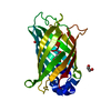
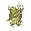
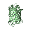
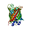
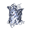
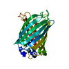
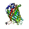
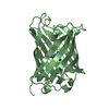
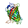
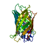
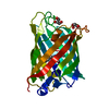
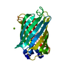
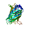
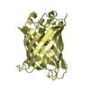
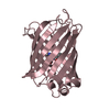
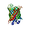
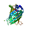
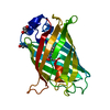
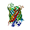
- PDB-1f0b: CRYSTAL STRUCTURE OF THE GREEN FLUORESCENT PROTEIN (GFP) VARIANT ... -
+ Open data

- Basic information
| Entry | Database: PDB / ID: 1f0b | |||||||||
|---|---|---|---|---|---|---|---|---|---|---|
| Title | CRYSTAL STRUCTURE OF THE GREEN FLUORESCENT PROTEIN (GFP) VARIANT YFP-H148Q | |||||||||
 Components Components | GREEN FLUORESCENT PROTEIN | |||||||||
 Keywords Keywords | LUMINESCENT PROTEIN / beta barrel / luminescence / bioluminescence / photoactive protein / green fluorescent protein | |||||||||
| Function / homology |  Function and homology information Function and homology information | |||||||||
| Biological species |   Aequorea victoria (jellyfish) Aequorea victoria (jellyfish) | |||||||||
| Method |  X-RAY DIFFRACTION / Resolution: 2.1 Å X-RAY DIFFRACTION / Resolution: 2.1 Å | |||||||||
 Authors Authors | Wachter, R.M. / Yarbrough, D. / Kallio, K. / Remington, S.J. | |||||||||
 Citation Citation | Journal: J.Mol.Biol. / Year: 2000 Title: Crystallographic and energetic analysis of binding of selected anions to the yellow variants of green fluorescent protein. Authors: Wachter, R.M. / Yarbrough, D. / Kallio, K. / Remington, S.J. #1: Journal: CURR.BIOL. / Year: 1999Title: Sensitivity of the Yellow Variant of Green Fluorescent Protein to Halides and Nitrate Authors: Wachter, R.M. / Remington, S.J. | |||||||||
| History |
|
- Structure visualization
| Structure viewer | Molecule: MolmilJmol/JSmol |
|---|
- Downloads & links
-Download
| PDBx/mmCIF format | 1f0b.cif.gz | 61.6 KB | Display | PDBx/mmCIF format |
|---|---|---|---|---|
| PDB format | pdb1f0b.ent.gz | 44.2 KB | Display | PDB format |
| PDBx/mmJSON format | 1f0b.json.gz | Tree view | PDBx/mmJSON format | |
| Others |  Other downloads Other downloads |
-Validation report
| Arichive directory | https://data.pdbj.org/pub/pdb/validation_reports/f0/1f0bftp://data.pdbj.org/pub/pdb/validation_reports/f0/1f0b | HTTPS FTP |
|---|
-Related structure data


-Links
 PDBj
PDBj

- Assembly
| Deposited unit | 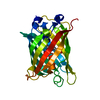
| ||||||||
|---|---|---|---|---|---|---|---|---|---|
| 1 |
| ||||||||
| Unit cell |
|
-Components
| #1: Protein | Mass: 26951.408 Da / Num. of mol.: 1 / Mutation: S65G, V68L, S72A, Q80R, T203Y, H148Q Source method: isolated from a genetically manipulated source Source: (gene. exp.) Aequorea victoria (jellyfish) / Plasmid: BL21DE3 / Production host:  |
|---|---|
| #2: Water | ChemComp-HOH /  Mass: 18.015 Da / Num. of mol.: 125 / Source method: isolated from a natural source / Formula: H2O Mass: 18.015 Da / Num. of mol.: 125 / Source method: isolated from a natural source / Formula: H2O |
| Has protein modification | Y |
-Experimental details
-Experiment
| Experiment | Method: X-RAY DIFFRACTION / Number of used crystals: 1 |
|---|
- Sample preparation
| Crystal | Density Matthews: 1.98 Å3/Da / Density % sol: 38 % | ||||||||||||||||||||||||||||||||||||
|---|---|---|---|---|---|---|---|---|---|---|---|---|---|---|---|---|---|---|---|---|---|---|---|---|---|---|---|---|---|---|---|---|---|---|---|---|---|
| Crystal grow | Temperature: 277 K / Method: vapor diffusion, hanging drop / pH: 5.5 Details: PEG 1550, sodium acetate, magnesium chloride, pH 5.5, VAPOR DIFFUSION, HANGING DROP, temperature 277.0K | ||||||||||||||||||||||||||||||||||||
| Crystal grow | *PLUS Temperature: 4 ℃ / pH: 7.9 | ||||||||||||||||||||||||||||||||||||
| Components of the solutions | *PLUS
|
-Data collection
| Diffraction | Mean temperature: 100 K |
|---|---|
| Diffraction source | Source: ROTATING ANODE / Type: RIGAKU / Wavelength: 1.5418 |
| Detector | Type: RIGAKU RAXIS IIC / Detector: IMAGE PLATE / Date: Oct 4, 1999 |
| Radiation | Protocol: SINGLE WAVELENGTH / Monochromatic (M) / Laue (L): M / Scattering type: x-ray |
| Radiation wavelength | Wavelength: 1.5418 Å / Relative weight: 1 |
| Reflection | Resolution: 2.07→22.5 Å / Num. all: 29297 / Num. obs: 11419 / % possible obs: 82.1 % / Redundancy: 2.56 % / Biso Wilson estimate: 25.3 Å2 / Rmerge(I) obs: 0.083 / Net I/σ(I): 10.3 |
| Reflection shell | Resolution: 2.07→2.15 Å / Redundancy: 2.5 % / Rmerge(I) obs: 0.32 / Num. unique all: 1106 / % possible all: 81.5 |
| Reflection | *PLUS Num. measured all: 29297 |
| Reflection shell | *PLUS % possible obs: 81.5 % |
- Processing
| Software |
| ||||||||||||||||
|---|---|---|---|---|---|---|---|---|---|---|---|---|---|---|---|---|---|
| Refinement | Resolution: 2.1→22.5 Å / Stereochemistry target values: Engh & Huber
| ||||||||||||||||
| Refinement step | Cycle: LAST / Resolution: 2.1→22.5 Å
| ||||||||||||||||
| Refine LS restraints |
| ||||||||||||||||
| Software | *PLUS Name: TNT / Classification: refinement | ||||||||||||||||
| Refinement | *PLUS Rfactor all: 0.188 | ||||||||||||||||
| Solvent computation | *PLUS | ||||||||||||||||
| Displacement parameters | *PLUS | ||||||||||||||||
| Refine LS restraints | *PLUS Type: t_angle_deg / Dev ideal: 3.06 |
