 Movie
Movie Controller

[English] 日本語
 Yorodumi
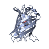
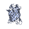
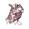
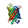
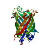
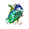
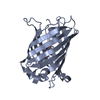
- PDB-1qyf: Crystal structure of matured green fluorescent protein R96A variant -
+ Open data

- Basic information
| Entry | Database: PDB / ID: 1qyf | |||||||||
|---|---|---|---|---|---|---|---|---|---|---|
| Title | Crystal structure of matured green fluorescent protein R96A variant | |||||||||
 Components Components | green-fluorescent protein | |||||||||
 Keywords Keywords | LUMINESCENT PROTEIN / beta barrel / chromophore | |||||||||
| Function / homology |  Function and homology information Function and homology information | |||||||||
| Biological species |   Aequorea victoria (jellyfish) Aequorea victoria (jellyfish) | |||||||||
| Method |  X-RAY DIFFRACTION / SYNCHROTRON / MOLECULAR REPLACEMENT / Resolution: 1.5 Å X-RAY DIFFRACTION / SYNCHROTRON / MOLECULAR REPLACEMENT / Resolution: 1.5 Å | |||||||||
 Authors Authors | Barondeau, D.P. / Putnam, C.D. / Kassmann, C.J. / Tainer, J.A. / Getzoff, E.D. | |||||||||
 Citation Citation | Journal: Proc.Natl.Acad.Sci.Usa / Year: 2003 Title: Mechanism and energetics of green fluorescent protein chromophore synthesis revealed by trapped intermediate structures. Authors: Barondeau, D.P. / Putnam, C.D. / Kassmann, C.J. / Tainer, J.A. / Getzoff, E.D. | |||||||||
| History |
|
- Structure visualization
| Structure viewer | Molecule: MolmilJmol/JSmol |
|---|
- Downloads & links
-Download
| PDBx/mmCIF format | 1qyf.cif.gz | 116.4 KB | Display | PDBx/mmCIF format |
|---|---|---|---|---|
| PDB format | pdb1qyf.ent.gz | 88.3 KB | Display | PDB format |
| PDBx/mmJSON format | 1qyf.json.gz | Tree view | PDBx/mmJSON format | |
| Others |  Other downloads Other downloads |
-Validation report
| Arichive directory | https://data.pdbj.org/pub/pdb/validation_reports/qy/1qyfftp://data.pdbj.org/pub/pdb/validation_reports/qy/1qyf | HTTPS FTP |
|---|
-Related structure data
| Related structure data |  1qxtC  1qy3C 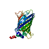 1qyoC  1qyqC  1emaS C: citing same article ( S: Starting model for refinement |
|---|---|
| Similar structure data |


-Links
 PDBj
PDBj

- Assembly
| Deposited unit | 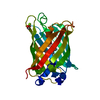
| ||||||||
|---|---|---|---|---|---|---|---|---|---|
| 1 |
| ||||||||
| Unit cell |
|
-Components
| #1: Protein | Mass: 25671.809 Da / Num. of mol.: 1 / Fragment: residues 1-229 Mutation: R96A, F99S, M153T, V163A, F64L, S65CRO, Y66CRO, G67CRO Source method: isolated from a genetically manipulated source Source: (gene. exp.) Aequorea victoria (jellyfish) / Gene: GFP / Production host:  |
|---|---|
| #2: Chemical | ChemComp-MG /   Mass: 24.305 Da / Num. of mol.: 1 / Source method: obtained synthetically / Formula: Mg Mass: 24.305 Da / Num. of mol.: 1 / Source method: obtained synthetically / Formula: Mg |
| #3: Chemical | ChemComp-EDO /   Mass: 62.068 Da / Num. of mol.: 1 / Source method: obtained synthetically / Formula: C2H6O2 Mass: 62.068 Da / Num. of mol.: 1 / Source method: obtained synthetically / Formula: C2H6O2 |
| #4: Water | ChemComp-HOH /  Mass: 18.015 Da / Num. of mol.: 275 / Source method: isolated from a natural source / Formula: H2O Mass: 18.015 Da / Num. of mol.: 275 / Source method: isolated from a natural source / Formula: H2O |
| Has protein modification | Y |
-Experimental details
-Experiment
| Experiment | Method: X-RAY DIFFRACTION |
|---|
- Sample preparation
| Crystal | Density Matthews: 2.22 Å3/Da / Density % sol: 44.68 % |
|---|---|
| Crystal grow | Temperature: 298 K / Method: vapor diffusion, hanging drop / pH: 8 Details: PEG 4000, Magnesium Chloride, Hepes, pH 8.0, VAPOR DIFFUSION, HANGING DROP, temperature 298K |
-Data collection
| Diffraction | Mean temperature: 100 K |
|---|---|
| Diffraction source | Source: SYNCHROTRON / Site: APS  / Beamline: 14-BM-C / Wavelength: 1 Å / Beamline: 14-BM-C / Wavelength: 1 Å |
| Detector | Type: ADSC QUANTUM 4 / Detector: CCD / Date: Sep 27, 1999 |
| Radiation | Protocol: SINGLE WAVELENGTH / Monochromatic (M) / Laue (L): M / Scattering type: x-ray |
| Radiation wavelength | Wavelength: 1 Å / Relative weight: 1 |
| Reflection | Resolution: 1.5→40 Å / Num. all: 37602 / Num. obs: 34756 / % possible obs: 92.4 % / Observed criterion σ(F): -3 / Observed criterion σ(I): -3 / Redundancy: 4.74 % / Biso Wilson estimate: 15.8 Å2 / Rsym value: 0.052 / Net I/σ(I): 27.4 |
| Reflection shell | Resolution: 1.5→1.55 Å / Mean I/σ(I) obs: 3 / Rsym value: 0.253 / % possible all: 60.7 |
- Processing
| Software |
| |||||||||||||||||||||||||||||||||
|---|---|---|---|---|---|---|---|---|---|---|---|---|---|---|---|---|---|---|---|---|---|---|---|---|---|---|---|---|---|---|---|---|---|---|
| Refinement | Method to determine structure: MOLECULAR REPLACEMENT Starting model: 1ema Resolution: 1.5→40 Å / Num. parameters: 18771 / Num. restraintsaints: 22556 / Cross valid method: FREE R / σ(F): 0 / Stereochemistry target values: ENGH AND HUBER Details: ANISOTROPIC REFINEMENT REDUCED FREE R (NO CUTOFF) BY 0.0196
| |||||||||||||||||||||||||||||||||
| Refine analyze | Num. disordered residues: 0 / Occupancy sum hydrogen: 1737 / Occupancy sum non hydrogen: 2084 | |||||||||||||||||||||||||||||||||
| Refinement step | Cycle: LAST / Resolution: 1.5→40 Å
| |||||||||||||||||||||||||||||||||
| Refine LS restraints |
|
