 Movie
Movie Controller

[English] 日本語
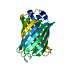
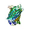
 Yorodumi
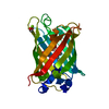
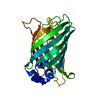
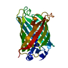
- PDB-2g2s: Structure of S65G Y66S GFP variant after spontaneous peptide hydr... -
+ Open data

- Basic information
| Entry | Database: PDB / ID: 2g2s | ||||||
|---|---|---|---|---|---|---|---|
| Title | Structure of S65G Y66S GFP variant after spontaneous peptide hydrolysis | ||||||
 Components Components | (Green fluorescent protein) x 2 | ||||||
 Keywords Keywords | LUMINESCENT PROTEIN / chromophore / biosynthesis / dehydroalanine / peptide hydrolysis / post-translational modification | ||||||
| Function / homology |  Function and homology information Function and homology information | ||||||
| Biological species |   Aequorea victoria (jellyfish) Aequorea victoria (jellyfish) | ||||||
| Method |  X-RAY DIFFRACTION / SYNCHROTRON / MOLECULAR REPLACEMENT / Resolution: 1.2 Å X-RAY DIFFRACTION / SYNCHROTRON / MOLECULAR REPLACEMENT / Resolution: 1.2 Å | ||||||
 Authors Authors | Barondeau, D.P. | ||||||
 Citation Citation | Journal: J.Am.Chem.Soc. / Year: 2006 Title: Understanding GFP Posttranslational Chemistry: Structures of Designed Variants that Achieve Backbone Fragmentation, Hydrolysis, and Decarboxylation. Authors: Barondeau, D.P. / Kassmann, C.J. / Tainer, J.A. / Getzoff, E.D. | ||||||
| History |
| ||||||
| Remark 999 | SEQUENCE Residue Ser 66, which is a Y66S mutation undergoes dehydration resulting in dehydroalanine moiety |
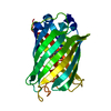
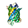
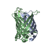
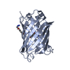
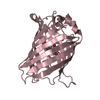
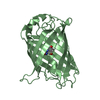
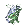
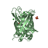
- Structure visualization
| Structure viewer | Molecule: MolmilJmol/JSmol |
|---|
- Downloads & links
-Download
| PDBx/mmCIF format | 2g2s.cif.gz | 68.6 KB | Display | PDBx/mmCIF format |
|---|---|---|---|---|
| PDB format | pdb2g2s.ent.gz | 48.1 KB | Display | PDB format |
| PDBx/mmJSON format | 2g2s.json.gz | Tree view | PDBx/mmJSON format | |
| Others |  Other downloads Other downloads |
-Validation report
| Arichive directory | https://data.pdbj.org/pub/pdb/validation_reports/g2/2g2sftp://data.pdbj.org/pub/pdb/validation_reports/g2/2g2s | HTTPS FTP |
|---|
-Related structure data
| Related structure data |  2g16C 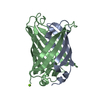 2g3dC  2g5zC  2g6eC  1emaS C: citing same article ( S: Starting model for refinement |
|---|---|
| Similar structure data |


-Links
 PDBj
PDBj

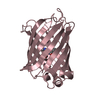
- Assembly
| Deposited unit | 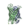
| ||||||||
|---|---|---|---|---|---|---|---|---|---|
| 1 |
| ||||||||
| Unit cell |
|
-Components
| #1: Protein | Mass: 6928.929 Da / Num. of mol.: 1 / Mutation: F64L, S65G Source method: isolated from a genetically manipulated source Source: (gene. exp.) Aequorea victoria (jellyfish) / Gene: GFP / Plasmid: pET11a / Species (production host): Escherichia coli / Production host:  |
|---|---|
| #2: Protein | Mass: 19806.074 Da / Num. of mol.: 1 / Mutation: Y66S, F99S, M153T, V163A Source method: isolated from a genetically manipulated source Source: (gene. exp.) Aequorea victoria (jellyfish) / Gene: GFP / Plasmid: pET11a / Species (production host): Escherichia coli / Production host: |
| #3: Chemical | ChemComp-MG /   Mass: 24.305 Da / Num. of mol.: 1 / Source method: obtained synthetically / Formula: Mg Mass: 24.305 Da / Num. of mol.: 1 / Source method: obtained synthetically / Formula: Mg |
| #4: Water | ChemComp-HOH /  Mass: 18.015 Da / Num. of mol.: 325 / Source method: isolated from a natural source / Formula: H2O Mass: 18.015 Da / Num. of mol.: 325 / Source method: isolated from a natural source / Formula: H2O |
-Experimental details
-Experiment
| Experiment | Method: X-RAY DIFFRACTION / Number of used crystals: 1 |
|---|
- Sample preparation
| Crystal | Density Matthews: 2.03 Å3/Da / Density % sol: 39.54 % |
|---|---|
| Crystal grow | Temperature: 298 K / pH: 8 Details: 50 mM MgCl2, 50 mM Hepes, 20% PEG 4000, pH 8.0, VAPOR DIFFUSION, HANGING DROP, temperature 298K, pH 8.00 |
-Data collection
| Diffraction | Mean temperature: 100 K |
|---|---|
| Diffraction source | Source: SYNCHROTRON / Site: SSRL  / Beamline: BL11-1 / Wavelength: 0.95369 / Beamline: BL11-1 / Wavelength: 0.95369 |
| Detector | Type: ADSC QUANTUM 315 / Detector: CCD / Date: Jan 20, 2003 |
| Radiation | Protocol: SINGLE WAVELENGTH / Monochromatic (M) / Laue (L): M / Scattering type: x-ray |
| Radiation wavelength | Wavelength: 0.95369 Å / Relative weight: 1 |
| Reflection | Resolution: 1.2→17.8 Å / Num. obs: 68670 / % possible obs: 99.3 % / Observed criterion σ(I): -3 / Biso Wilson estimate: 11 Å2 / Rsym value: 0.044 / Net I/σ(I): 44 |
| Reflection shell | Resolution: 1.2→1.24 Å / Mean I/σ(I) obs: 5.1 / Rsym value: 0.292 / % possible all: 94.7 |
- Processing
| Software |
| |||||||||||||||||||||||||||||||||
|---|---|---|---|---|---|---|---|---|---|---|---|---|---|---|---|---|---|---|---|---|---|---|---|---|---|---|---|---|---|---|---|---|---|---|
| Refinement | Method to determine structure: MOLECULAR REPLACEMENT Starting model: PDB ENTRY 1EMA Resolution: 1.2→17.8 Å / Cross valid method: THROUGHOUT / σ(F): 0 / Stereochemistry target values: Engh & Huber
| |||||||||||||||||||||||||||||||||
| Refinement step | Cycle: LAST / Resolution: 1.2→17.8 Å
| |||||||||||||||||||||||||||||||||
| Refine LS restraints |
|
