 Movie
Movie Controller

[English] 日本語
 Yorodumi
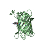
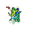
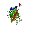
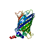
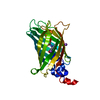
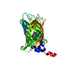
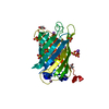
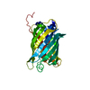
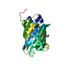
- PDB-2hcg: Structure of S65T Y66F GFP variant after cyclization, carbon-carb... -
+ Open data

- Basic information
| Entry | Database: PDB / ID: 2hcg | |||||||||
|---|---|---|---|---|---|---|---|---|---|---|
| Title | Structure of S65T Y66F GFP variant after cyclization, carbon-carbon bond cleavage, and oxygen incorporation reactions | |||||||||
 Components Components | Green fluorescent protein | |||||||||
 Keywords Keywords | LUMINESCENT PROTEIN / post-translational modification / cyclization / cleavage / fluorophore | |||||||||
| Function / homology |  Function and homology information Function and homology information | |||||||||
| Biological species |   Aequorea victoria (jellyfish) Aequorea victoria (jellyfish) | |||||||||
| Method |  X-RAY DIFFRACTION / SYNCHROTRON / MOLECULAR REPLACEMENT / Resolution: 1.35 Å X-RAY DIFFRACTION / SYNCHROTRON / MOLECULAR REPLACEMENT / Resolution: 1.35 Å | |||||||||
 Authors Authors | Barondeau, D.P. / Kassmann, C.J. / Tainer, J.A. / Getzoff, E.D. | |||||||||
 Citation Citation | Journal: J.Am.Chem.Soc. / Year: 2007 Title: The Case of the Missing Ring: Radical Cleavage of a Carbon-Carbon Bond and Implications for GFP Chromophore Biosynthesis Authors: Barondeau, D.P. / Kassmann, C.J. / Tainer, J.A. / Getzoff, E.D. | |||||||||
| History |
|
- Structure visualization
| Structure viewer | Molecule: MolmilJmol/JSmol |
|---|
- Downloads & links
-Download
| PDBx/mmCIF format | 2hcg.cif.gz | 71.2 KB | Display | PDBx/mmCIF format |
|---|---|---|---|---|
| PDB format | pdb2hcg.ent.gz | 50.3 KB | Display | PDB format |
| PDBx/mmJSON format | 2hcg.json.gz | Tree view | PDBx/mmJSON format | |
| Others |  Other downloads Other downloads |
-Validation report
| Arichive directory | https://data.pdbj.org/pub/pdb/validation_reports/hc/2hcgftp://data.pdbj.org/pub/pdb/validation_reports/hc/2hcg | HTTPS FTP |
|---|
-Related structure data
| Related structure data |  2hfcC  2hgdC  2hgyC  1emaS S: Starting model for refinement C: citing same article ( |
|---|---|
| Similar structure data |

-Links
 PDBj
PDBj

- Assembly
| Deposited unit | 
| ||||||||
|---|---|---|---|---|---|---|---|---|---|
| 1 |
| ||||||||
| Unit cell |
|
-Components
| #1: Protein | Mass: 26763.029 Da / Num. of mol.: 1 / Mutation: F64L, S65T, Y66F, F99S, M153T, V163A Source method: isolated from a genetically manipulated source Source: (gene. exp.) Aequorea victoria (jellyfish) / Gene: GFP / Plasmid: pET11a / Production host:  |
|---|---|
| #2: Chemical | ChemComp-MG /   Mass: 24.305 Da / Num. of mol.: 1 / Source method: obtained synthetically / Formula: Mg Mass: 24.305 Da / Num. of mol.: 1 / Source method: obtained synthetically / Formula: Mg |
| #3: Water | ChemComp-HOH /  Mass: 18.015 Da / Num. of mol.: 399 / Source method: isolated from a natural source / Formula: H2O Mass: 18.015 Da / Num. of mol.: 399 / Source method: isolated from a natural source / Formula: H2O |
| Has protein modification | Y |
| Sequence details | RESIDUES THR 65, PHE 66 AND GLY 67 CONSTITUTE THE CHROMOPHORE C99. RESIDUE AT POSITION 65 IS S65T ...RESIDUES THR 65, PHE 66 AND GLY 67 CONSTITUTE |
-Experimental details
-Experiment
| Experiment | Method: X-RAY DIFFRACTION / Number of used crystals: 1 |
|---|
- Sample preparation
| Crystal | Density Matthews: 2.11 Å3/Da / Density % sol: 41.8 % |
|---|---|
| Crystal grow | Temperature: 298 K / Method: vapor diffusion, hanging drop / pH: 8 Details: 19% PEG 4K, 50 mM MgCl2, 50 mM Hepes, pH 8.0, VAPOR DIFFUSION, HANGING DROP, temperature 298K |
-Data collection
| Diffraction | Mean temperature: 100 K |
|---|---|
| Diffraction source | Source: SYNCHROTRON / Site: SSRL  / Beamline: BL9-1 / Wavelength: 0.9706 Å / Beamline: BL9-1 / Wavelength: 0.9706 Å |
| Detector | Type: MAR scanner 345 mm plate / Detector: IMAGE PLATE / Date: Dec 7, 2001 |
| Radiation | Protocol: SINGLE WAVELENGTH / Monochromatic (M) / Laue (L): M / Scattering type: x-ray |
| Radiation wavelength | Wavelength: 0.9706 Å / Relative weight: 1 |
| Reflection | Resolution: 1.35→31 Å / Num. obs: 50300 / % possible obs: 98.9 % / Observed criterion σ(F): 0 / Observed criterion σ(I): -3 / Biso Wilson estimate: 11.6 Å2 / Rsym value: 0.043 / Net I/σ(I): 26.2 |
| Reflection shell | Resolution: 1.35→1.4 Å / Rmerge(I) obs: 0.352 / Mean I/σ(I) obs: 3.1 / Num. unique all: 4869 / % possible all: 97.2 |
- Processing
| Software |
| ||||||||||||||||||||
|---|---|---|---|---|---|---|---|---|---|---|---|---|---|---|---|---|---|---|---|---|---|
| Refinement | Method to determine structure: MOLECULAR REPLACEMENT Starting model: PDB ENTRY 1ema Resolution: 1.35→31 Å / Cross valid method: THROUGHOUT / σ(F): 0 / σ(I): 0 / Stereochemistry target values: Engh & Huber
| ||||||||||||||||||||
| Refinement step | Cycle: LAST / Resolution: 1.35→31 Å
| ||||||||||||||||||||
| Refine LS restraints |
|
