 Movie
Movie Controller


+ Open data

- Basic information
| Entry | Database: PDB / ID: 3w1c | |||||||||
|---|---|---|---|---|---|---|---|---|---|---|
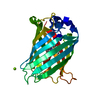
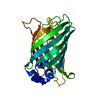
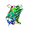
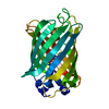
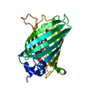
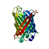
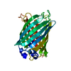
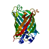
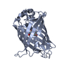
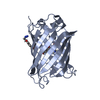
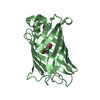
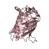
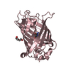
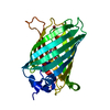
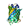
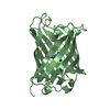
| Title | Structure of a pressure sensitive YFP variant YFP-G1 | |||||||||
 Components Components | Green Fluorescent protein | |||||||||
 Keywords Keywords | LUMINESCENT PROTEIN / beta barrel / fluorescent protein | |||||||||
| Function / homology |  Function and homology information Function and homology information | |||||||||
| Biological species |   Aequorea victoria (jellyfish) Aequorea victoria (jellyfish) | |||||||||
| Method |  X-RAY DIFFRACTION / SYNCHROTRON / MOLECULAR REPLACEMENT / Resolution: 1.3 Å X-RAY DIFFRACTION / SYNCHROTRON / MOLECULAR REPLACEMENT / Resolution: 1.3 Å | |||||||||
 Authors Authors | Imada, K. / Yoshizawa, K. / Kinoshita, M. / Watanabe, T.M. | |||||||||
 Citation Citation | Journal: Plos One / Year: 2013 Title: Glycine insertion makes yellow fluorescent protein sensitive to hydrostatic pressure. Authors: Watanabe, T.M. / Imada, K. / Yoshizawa, K. / Nishiyama, M. / Kato, C. / Abe, F. / Morikawa, T.J. / Kinoshita, M. / Fujita, H. / Yanagida, T. | |||||||||
| History |
|
- Structure visualization
| Structure viewer | Molecule: MolmilJmol/JSmol |
|---|
- Downloads & links
-Download
| PDBx/mmCIF format | 3w1c.cif.gz | 68.6 KB | Display | PDBx/mmCIF format |
|---|---|---|---|---|
| PDB format | pdb3w1c.ent.gz | 48.8 KB | Display | PDB format |
| PDBx/mmJSON format | 3w1c.json.gz | Tree view | PDBx/mmJSON format | |
| Others |  Other downloads Other downloads |
-Validation report
| Arichive directory | https://data.pdbj.org/pub/pdb/validation_reports/w1/3w1cftp://data.pdbj.org/pub/pdb/validation_reports/w1/3w1c | HTTPS FTP |
|---|
-Related structure data
| Related structure data |  3w1dC 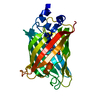 3dq7S C: citing same article ( S: Starting model for refinement |
|---|---|
| Similar structure data |


-Links
 PDBj
PDBj

- Assembly
| Deposited unit | 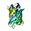
| ||||||||
|---|---|---|---|---|---|---|---|---|---|
| 1 |
| ||||||||
| Unit cell |
|
-Components
| #1: Protein | Mass: 26932.359 Da / Num. of mol.: 1 / Mutation: S65G, V68L, S72A, T204Y, H232L Source method: isolated from a genetically manipulated source Source: (gene. exp.) Aequorea victoria (jellyfish) / Gene: GFP / Plasmid: pAL7 / Production host:  |
|---|---|
| #2: Water | ChemComp-HOH /  Mass: 18.015 Da / Num. of mol.: 410 / Source method: isolated from a natural source / Formula: H2O Mass: 18.015 Da / Num. of mol.: 410 / Source method: isolated from a natural source / Formula: H2O |
| Has protein modification | Y |
| Sequence details | RESIDUE SER 65 HAS BEEN MUTATED TO GLY 65. RESIDUES GLY 65, TYR 66 AND GLY 67 CONSTITUTE THE ...RESIDUE SER 65 HAS BEEN MUTATED TO GLY 65. RESIDUES GLY 65, TYR 66 AND GLY 67 CONSTITUTE |
-Experimental details
-Experiment
| Experiment | Method: X-RAY DIFFRACTION / Number of used crystals: 1 |
|---|
- Sample preparation
| Crystal | Density Matthews: 2.08 Å3/Da / Density % sol: 40.94 % |
|---|---|
| Crystal grow | Temperature: 277 K / Method: vapor diffusion, sitting drop / pH: 6.5 Details: 13-15% PEG 8000, 0.1M Bis-Tris buffer, 350-400mM calcium acetate , pH 6.5, VAPOR DIFFUSION, SITTING DROP, temperature 277K |
-Data collection
| Diffraction | Mean temperature: 100 K |
|---|---|
| Diffraction source | Source: SYNCHROTRON / Site: SPring-8  / Beamline: BL38B1 / Wavelength: 1 Å / Beamline: BL38B1 / Wavelength: 1 Å |
| Detector | Type: ADSC QUANTUM 210 / Detector: CCD / Date: Apr 12, 2010 |
| Radiation | Monochromator: Double-crystal monochromator / Protocol: SINGLE WAVELENGTH / Monochromatic (M) / Laue (L): M / Scattering type: x-ray |
| Radiation wavelength | Wavelength: 1 Å / Relative weight: 1 |
| Reflection | Resolution: 1.3→17.3 Å / Num. all: 54434 / Num. obs: 54434 / % possible obs: 97.3 % / Observed criterion σ(F): 0 / Observed criterion σ(I): 0 / Redundancy: 5.6 % / Biso Wilson estimate: 11.1 Å2 / Rmerge(I) obs: 0.065 / Rsym value: 0.065 / Net I/σ(I): 15.9 |
| Reflection shell | Resolution: 1.3→1.37 Å / Redundancy: 5.3 % / Rmerge(I) obs: 0.469 / Mean I/σ(I) obs: 3.5 / Num. unique all: 7655 / % possible all: 94.5 |
- Processing
| Software |
| ||||||||||||||||||||||||||||||||||||||||||||||||||||||||||||||||||||||||||||||||
|---|---|---|---|---|---|---|---|---|---|---|---|---|---|---|---|---|---|---|---|---|---|---|---|---|---|---|---|---|---|---|---|---|---|---|---|---|---|---|---|---|---|---|---|---|---|---|---|---|---|---|---|---|---|---|---|---|---|---|---|---|---|---|---|---|---|---|---|---|---|---|---|---|---|---|---|---|---|---|---|---|---|
| Refinement | Method to determine structure: MOLECULAR REPLACEMENT Starting model: PDB ENTRY 3DQ7 Resolution: 1.3→17.25 Å / Rfactor Rfree error: 0.004 / Data cutoff high absF: 1133833.32 / Data cutoff low absF: 0 / Isotropic thermal model: RESTRAINED / Cross valid method: THROUGHOUT / σ(F): 0 / Stereochemistry target values: Engh & Huber / Details: BULK SOLVENT MODEL USED
| ||||||||||||||||||||||||||||||||||||||||||||||||||||||||||||||||||||||||||||||||
| Solvent computation | Solvent model: FLAT MODEL / Bsol: 44.3911 Å2 / ksol: 0.45 e/Å3 | ||||||||||||||||||||||||||||||||||||||||||||||||||||||||||||||||||||||||||||||||
| Displacement parameters | Biso mean: 14.8 Å2
| ||||||||||||||||||||||||||||||||||||||||||||||||||||||||||||||||||||||||||||||||
| Refine analyze |
| ||||||||||||||||||||||||||||||||||||||||||||||||||||||||||||||||||||||||||||||||
| Refinement step | Cycle: LAST / Resolution: 1.3→17.25 Å
| ||||||||||||||||||||||||||||||||||||||||||||||||||||||||||||||||||||||||||||||||
| Refine LS restraints |
| ||||||||||||||||||||||||||||||||||||||||||||||||||||||||||||||||||||||||||||||||
| LS refinement shell | Resolution: 1.3→1.38 Å / Rfactor Rfree error: 0.013 / Total num. of bins used: 6
| ||||||||||||||||||||||||||||||||||||||||||||||||||||||||||||||||||||||||||||||||
| Xplor file |
|
