[English] 日本語
 Yorodumi
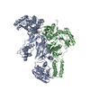
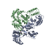
- PDB-2vg5: Crystal structures of HIV-1 reverse transcriptase complexes with ... -
+ Open data
- Basic information
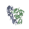
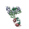
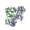
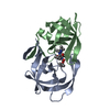
| Entry | Database: PDB / ID: 2vg5 | ||||||
|---|---|---|---|---|---|---|---|
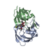
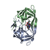
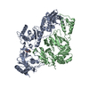
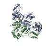
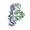
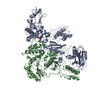
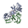
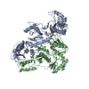
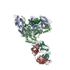
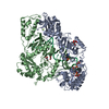
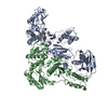
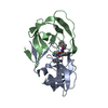
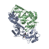
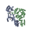
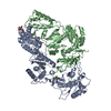
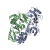
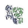
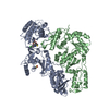
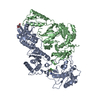
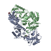
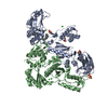
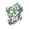
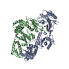
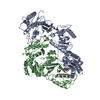
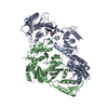
| Title | Crystal structures of HIV-1 reverse transcriptase complexes with thiocarbamate non-nucleoside inhibitors | ||||||
 Components Components |
| ||||||
 Keywords Keywords | TRANSFERASE / DNA-DIRECTED DNA POLYMERASE / THIOCARBAMATES / PHOSPHORYLATION / DNA INTEGRATION / MAGNESIUM / ZINC-FINGER / RNA-BINDING / LIPOPROTEIN / CORE PROTEIN / ENDONUCLEASE / METAL-BINDING / ZINC / AIDS / HIV-1 / VIRION / NUCLEUS / MEMBRANE / ASPARTYL PROTEASE / CAPSID MATURATION / MULTIFUNCTIONAL ENZYME / RNA-DIRECTED DNA POLYMERASE / REVERSE TRANSCRIPTASE / NUCLEOTIDYLTRANSFERASE / DNA RECOMBINATION / VIRAL NUCLEOPROTEIN / PROTEASE / NUCLEASE / MYRISTATE / HYDROLASE / CYTOPLASM / NON-NUCLEOSIDE REVERSE TRANSCRIPTASE INHIBITORS | ||||||
| Function / homology |  Function and homology information Function and homology informationHIV-1 retropepsin / symbiont-mediated activation of host apoptosis / retroviral ribonuclease H / exoribonuclease H / exoribonuclease H activity / DNA integration / viral genome integration into host DNA / establishment of integrated proviral latency / RNA-directed DNA polymerase / RNA stem-loop binding ...HIV-1 retropepsin / symbiont-mediated activation of host apoptosis / retroviral ribonuclease H / exoribonuclease H / exoribonuclease H activity / DNA integration / viral genome integration into host DNA / establishment of integrated proviral latency / RNA-directed DNA polymerase / RNA stem-loop binding / viral penetration into host nucleus / host multivesicular body / RNA-directed DNA polymerase activity / RNA-DNA hybrid ribonuclease activity / Transferases; Transferring phosphorus-containing groups; Nucleotidyltransferases / host cell / viral nucleocapsid / DNA recombination / DNA-directed DNA polymerase / aspartic-type endopeptidase activity / Hydrolases; Acting on ester bonds / DNA-directed DNA polymerase activity / symbiont-mediated suppression of host gene expression / viral translational frameshifting / symbiont entry into host cell / lipid binding / host cell nucleus / host cell plasma membrane / virion membrane / structural molecule activity / proteolysis / DNA binding / zinc ion binding Similarity search - Function | ||||||
| Biological species |   HUMAN IMMUNODEFICIENCY VIRUS 1 HUMAN IMMUNODEFICIENCY VIRUS 1 | ||||||
| Method |  X-RAY DIFFRACTION / X-RAY DIFFRACTION /  SYNCHROTRON / SYNCHROTRON /  MOLECULAR REPLACEMENT / Resolution: 2.8 Å MOLECULAR REPLACEMENT / Resolution: 2.8 Å | ||||||
 Authors Authors | Spallarossa, A. / Cesarini, S. / Ranise, A. / Ponassi, M. / Unge, T. / Bolognesi, M. | ||||||
 Citation Citation |  Journal: Biochem.Biophys.Res.Commun. / Year: 2008 Journal: Biochem.Biophys.Res.Commun. / Year: 2008Title: Crystal Structures of HIV-1 Reverse Transcriptase Complexes with Thiocarbamate Non-Nucleoside Inhibitors. Authors: Spallarossa, A. / Cesarini, S. / Ranise, A. / Ponassi, M. / Unge, T. / Bolognesi, M. | ||||||
| History |
| ||||||
| Remark 700 | SHEET THE SHEET STRUCTURE OF THIS MOLECULE IS BIFURCATED. IN ORDER TO REPRESENT THIS FEATURE IN ... SHEET THE SHEET STRUCTURE OF THIS MOLECULE IS BIFURCATED. IN ORDER TO REPRESENT THIS FEATURE IN THE SHEET RECORDS BELOW, TWO SHEETS ARE DEFINED. |
- Structure visualization
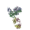
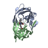
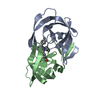
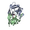
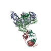
| Structure viewer | Molecule:  Molmil Molmil Jmol/JSmol Jmol/JSmol |
|---|
- Downloads & links
- Download
| PDBx/mmCIF format |  2vg5.cif.gz 2vg5.cif.gz | 205.3 KB | Display |  PDBx/mmCIF format PDBx/mmCIF format |
|---|---|---|---|---|
| PDB format |  pdb2vg5.ent.gz pdb2vg5.ent.gz | 163.8 KB | Display |  PDB format PDB format |
| PDBx/mmJSON format |  2vg5.json.gz 2vg5.json.gz | Tree view |  PDBx/mmJSON format PDBx/mmJSON format | |
| Others |  Other downloads Other downloads |
-Validation report
| Arichive directory |  https://data.pdbj.org/pub/pdb/validation_reports/vg/2vg5 https://data.pdbj.org/pub/pdb/validation_reports/vg/2vg5 ftp://data.pdbj.org/pub/pdb/validation_reports/vg/2vg5 ftp://data.pdbj.org/pub/pdb/validation_reports/vg/2vg5 | HTTPS FTP |
|---|
-Related structure data
- Links
Links
- Assembly
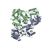
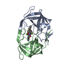
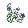
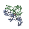
| Deposited unit | 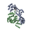
| ||||||||
|---|---|---|---|---|---|---|---|---|---|
| 1 |
| ||||||||
| Unit cell |
|
- Components
| #1: Protein | Mass: 64161.535 Da / Num. of mol.: 1 Fragment: GAG-POL POLYPROTEIN P66 SUBUNIT, RESIDUES 600-1156 Source method: isolated from a genetically manipulated source Source: (gene. exp.)   HUMAN IMMUNODEFICIENCY VIRUS 1 / Production host: HUMAN IMMUNODEFICIENCY VIRUS 1 / Production host:  References: UniProt: P03366, RNA-directed DNA polymerase, DNA-directed DNA polymerase, ribonuclease H |
|---|---|
| #2: Protein | Mass: 50055.551 Da / Num. of mol.: 1 Fragment: GAG-POL POLYPROTEIN P51 SUBUNIT, RESIDUES 600-1027 Source method: isolated from a genetically manipulated source Source: (gene. exp.)   HUMAN IMMUNODEFICIENCY VIRUS 1 / Production host: HUMAN IMMUNODEFICIENCY VIRUS 1 / Production host:  |
| #3: Chemical | ChemComp-NNC / |
| #4: Water | ChemComp-HOH / |
-Experimental details
-Experiment
| Experiment | Method:  X-RAY DIFFRACTION X-RAY DIFFRACTION |
|---|
- Sample preparation
| Crystal | Density Matthews: 3.25 Å3/Da / Density % sol: 61.9 % / Description: NONE |
|---|
-Data collection
| Diffraction | Mean temperature: 100 K |
|---|---|
| Diffraction source | Source:  SYNCHROTRON / Site: SYNCHROTRON / Site:  MAX II MAX II  / Beamline: I711 / Wavelength: 0.969 / Beamline: I711 / Wavelength: 0.969 |
| Detector | Type: MARRESEARCH / Detector: CCD |
| Radiation | Protocol: SINGLE WAVELENGTH / Monochromatic (M) / Laue (L): M / Scattering type: x-ray |
| Radiation wavelength | Wavelength: 0.969 Å / Relative weight: 1 |
| Reflection | Resolution: 2.8→20 Å / Num. obs: 31949 / % possible obs: 89.8 % / Observed criterion σ(I): 2 / Redundancy: 1.9 % / Rmerge(I) obs: 0.08 |
- Processing
| Software | Name: REFMAC / Version: 5.2.0005 / Classification: refinement | ||||||||||||||||||||||||||||||||||||||||||||||||||||||||||||||||||||||||||||||||||||||||||||||||||||||||||||||||||||||||||||||||||||||||||||||||||||||||||||||||||||||||||||||||||||||
|---|---|---|---|---|---|---|---|---|---|---|---|---|---|---|---|---|---|---|---|---|---|---|---|---|---|---|---|---|---|---|---|---|---|---|---|---|---|---|---|---|---|---|---|---|---|---|---|---|---|---|---|---|---|---|---|---|---|---|---|---|---|---|---|---|---|---|---|---|---|---|---|---|---|---|---|---|---|---|---|---|---|---|---|---|---|---|---|---|---|---|---|---|---|---|---|---|---|---|---|---|---|---|---|---|---|---|---|---|---|---|---|---|---|---|---|---|---|---|---|---|---|---|---|---|---|---|---|---|---|---|---|---|---|---|---|---|---|---|---|---|---|---|---|---|---|---|---|---|---|---|---|---|---|---|---|---|---|---|---|---|---|---|---|---|---|---|---|---|---|---|---|---|---|---|---|---|---|---|---|---|---|---|---|
| Refinement | Method to determine structure:  MOLECULAR REPLACEMENT / Resolution: 2.8→20 Å / Cor.coef. Fo:Fc: 0.911 / Cor.coef. Fo:Fc free: 0.854 / SU B: 16.484 / SU ML: 0.325 / Cross valid method: THROUGHOUT / ESU R Free: 0.449 / Stereochemistry target values: MAXIMUM LIKELIHOOD / Details: HYDROGENS HAVE BEEN ADDED IN THE RIDING POSITIONS. MOLECULAR REPLACEMENT / Resolution: 2.8→20 Å / Cor.coef. Fo:Fc: 0.911 / Cor.coef. Fo:Fc free: 0.854 / SU B: 16.484 / SU ML: 0.325 / Cross valid method: THROUGHOUT / ESU R Free: 0.449 / Stereochemistry target values: MAXIMUM LIKELIHOOD / Details: HYDROGENS HAVE BEEN ADDED IN THE RIDING POSITIONS.
| ||||||||||||||||||||||||||||||||||||||||||||||||||||||||||||||||||||||||||||||||||||||||||||||||||||||||||||||||||||||||||||||||||||||||||||||||||||||||||||||||||||||||||||||||||||||
| Solvent computation | Ion probe radii: 0.8 Å / Shrinkage radii: 0.8 Å / VDW probe radii: 1.2 Å / Solvent model: BABINET MODEL WITH MASK | ||||||||||||||||||||||||||||||||||||||||||||||||||||||||||||||||||||||||||||||||||||||||||||||||||||||||||||||||||||||||||||||||||||||||||||||||||||||||||||||||||||||||||||||||||||||
| Displacement parameters | Biso mean: 45.11 Å2
| ||||||||||||||||||||||||||||||||||||||||||||||||||||||||||||||||||||||||||||||||||||||||||||||||||||||||||||||||||||||||||||||||||||||||||||||||||||||||||||||||||||||||||||||||||||||
| Refinement step | Cycle: LAST / Resolution: 2.8→20 Å
| ||||||||||||||||||||||||||||||||||||||||||||||||||||||||||||||||||||||||||||||||||||||||||||||||||||||||||||||||||||||||||||||||||||||||||||||||||||||||||||||||||||||||||||||||||||||
| Refine LS restraints |
|
 Movie
Movie Controller
































 PDBj
PDBj


