 Movie
Movie Controller


+ Open data

- Basic information
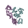
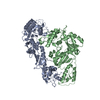
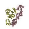
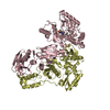
| Entry | Database: PDB / ID: 1dlo | ||||||
|---|---|---|---|---|---|---|---|
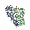
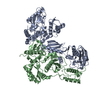
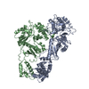
| Title | HUMAN IMMUNODEFICIENCY VIRUS TYPE 1 | ||||||
 Components Components | (HUMAN IMMUNODEFICIENCY VIRUS TYPE 1 REVERSE TRANSCRIPTASE) x 2 | ||||||
 Keywords Keywords | NUCLEOTIDYLTRANSFERASE | ||||||
| Function / homology |  Function and homology information Function and homology informationHIV-1 retropepsin / symbiont-mediated activation of host apoptosis / retroviral ribonuclease H / exoribonuclease H / exoribonuclease H activity / DNA integration / viral genome integration into host DNA / establishment of integrated proviral latency / RNA-directed DNA polymerase / RNA stem-loop binding ...HIV-1 retropepsin / symbiont-mediated activation of host apoptosis / retroviral ribonuclease H / exoribonuclease H / exoribonuclease H activity / DNA integration / viral genome integration into host DNA / establishment of integrated proviral latency / RNA-directed DNA polymerase / RNA stem-loop binding / viral penetration into host nucleus / host multivesicular body / RNA-directed DNA polymerase activity / RNA-DNA hybrid ribonuclease activity / Transferases; Transferring phosphorus-containing groups; Nucleotidyltransferases / host cell / viral nucleocapsid / DNA recombination / DNA-directed DNA polymerase / aspartic-type endopeptidase activity / Hydrolases; Acting on ester bonds / DNA-directed DNA polymerase activity / symbiont-mediated suppression of host gene expression / viral translational frameshifting / symbiont entry into host cell / lipid binding / host cell nucleus / host cell plasma membrane / virion membrane / structural molecule activity / proteolysis / DNA binding / zinc ion binding Similarity search - Function | ||||||
| Biological species |   Human immunodeficiency virus 1 Human immunodeficiency virus 1 | ||||||
| Method |  X-RAY DIFFRACTION / SYNCHROTRON / Resolution: 2.7 Å X-RAY DIFFRACTION / SYNCHROTRON / Resolution: 2.7 Å | ||||||
 Authors Authors | Hsiou, Y. / Ding, J. / Das, K. / Hughes, S. / Arnold, E. | ||||||
 Citation Citation | Journal: Structure / Year: 1996 Title: Structure of unliganded HIV-1 reverse transcriptase at 2.7 A resolution: implications of conformational changes for polymerization and inhibition mechanisms. Authors: Hsiou, Y. / Ding, J. / Das, K. / Clark Jr., A.D. / Hughes, S.H. / Arnold, E. #1: Journal: Structure / Year: 1995Title: Structure of HIV-1 Reverse Transcriptase in a Complex with the Non-Nucleoside Inhibitor Alpha-Apa R 95845 at 2.8 A Resolution Authors: Ding, J. / Das, K. / Tantillo, C. / Zhang, W. / Clark Junior, A.D. / Jessen, S. / Lu, X. / Hsiou, Y. / Jacobo-Molina, A. / Andries, K. / al., et #2: Journal: Nat.Struct.Biol. / Year: 1995Title: Structure of HIV-1 RT/TIBO R 86183 Complex Reveals Similarity in the Binding of Diverse Nonnucleoside Inhibitors Authors: Ding, J. / Das, K. / Moereels, H. / Koymans, L. / Andries, K. / Janssen, P.A.J. / Hughes, S.H. / Arnold, E. #3: Journal: Proc.Natl.Acad.Sci.USA / Year: 1993Title: Crystal Structure of Human Immunodeficiency Virus Type 1 Reverse Transcriptase Complexed with Double-Stranded DNA at 3.0 A Resolution Shows Bent DNA Authors: Jacobo-Molina, A. / Ding, J. / Nanni, R.G. / Clark Junior, A.D. / Lu, X. / Tantillo, C. / Williams, R.L. / Kamer, G. / Ferris, A.L. / Clark, P. / al., et | ||||||
| History |
|
- Structure visualization
| Structure viewer | Molecule: MolmilJmol/JSmol |
|---|
- Downloads & links
-Download
| PDBx/mmCIF format | 1dlo.cif.gz | 198.8 KB | Display | PDBx/mmCIF format |
|---|---|---|---|---|
| PDB format | pdb1dlo.ent.gz | 160.5 KB | Display | PDB format |
| PDBx/mmJSON format | 1dlo.json.gz | Tree view | PDBx/mmJSON format | |
| Others |  Other downloads Other downloads |
-Validation report
| Arichive directory | https://data.pdbj.org/pub/pdb/validation_reports/dl/1dloftp://data.pdbj.org/pub/pdb/validation_reports/dl/1dlo | HTTPS FTP |
|---|
-Related structure data
| Similar structure data |
|---|


-Links
 PDBj
PDBj


- Assembly
| Deposited unit | 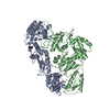
| ||||||||
|---|---|---|---|---|---|---|---|---|---|
| 1 |
| ||||||||
| Unit cell |
|
-Components
| #1: Protein | Mass: 63988.273 Da / Num. of mol.: 1 / Mutation: C280S Source method: isolated from a genetically manipulated source Source: (gene. exp.) Human immunodeficiency virus 1 / Genus: Lentivirus / Strain: BH10 ISOLATE / Production host:  |
|---|---|
| #2: Protein | Mass: 49911.359 Da / Num. of mol.: 1 / Mutation: C280S Source method: isolated from a genetically manipulated source Source: (gene. exp.) Human immunodeficiency virus 1 / Genus: Lentivirus / Strain: BH10 ISOLATE / Production host: |
| Compound details | HIV-1 RT IS COMPOSED OF TWO SUBUNITS OF 66 KDA AND 51 KDA, DESIGNATED AS P66 AND P51, RESPECTIVELY. ...HIV-1 RT IS COMPOSED OF TWO SUBUNITS OF 66 KDA AND 51 KDA, DESIGNATED |
-Experimental details
-Experiment
| Experiment | Method: X-RAY DIFFRACTION |
|---|
- Sample preparation
| Crystal | Density Matthews: 3.26 Å3/Da / Density % sol: 64 % | ||||||||||||||||||||||||||||||||||||||||||||||||||||||||||||||||||||||||||||||||||||
|---|---|---|---|---|---|---|---|---|---|---|---|---|---|---|---|---|---|---|---|---|---|---|---|---|---|---|---|---|---|---|---|---|---|---|---|---|---|---|---|---|---|---|---|---|---|---|---|---|---|---|---|---|---|---|---|---|---|---|---|---|---|---|---|---|---|---|---|---|---|---|---|---|---|---|---|---|---|---|---|---|---|---|---|---|---|
| Crystal | *PLUS | ||||||||||||||||||||||||||||||||||||||||||||||||||||||||||||||||||||||||||||||||||||
| Crystal grow | *PLUS pH: 7 / Method: vapor diffusion / Details: Kohlstaedt, L.A., (1992) Science, 256, 1783. | ||||||||||||||||||||||||||||||||||||||||||||||||||||||||||||||||||||||||||||||||||||
| Components of the solutions | *PLUS
|
-Data collection
| Diffraction source | Source: SYNCHROTRON / Site: CHESS  / Beamline: F1 / Wavelength: 0.908 / Beamline: F1 / Wavelength: 0.908 |
|---|---|
| Detector | Detector: IMAGE PLATE AREA DETECTOR / Date: Oct 1, 1994 |
| Radiation | Monochromatic (M) / Laue (L): M / Scattering type: x-ray |
| Radiation wavelength | Wavelength: 0.908 Å / Relative weight: 1 |
| Reflection | Num. obs: 41049 / % possible obs: 99.5 % / Redundancy: 3.5 % / Rmerge(I) obs: 0.055 |
| Reflection | *PLUS Highest resolution: 2.7 Å / Num. measured all: 142770 |
- Processing
| Software |
| ||||||||||||||||||||||||||||||||||||||||||||||||||||||||||||
|---|---|---|---|---|---|---|---|---|---|---|---|---|---|---|---|---|---|---|---|---|---|---|---|---|---|---|---|---|---|---|---|---|---|---|---|---|---|---|---|---|---|---|---|---|---|---|---|---|---|---|---|---|---|---|---|---|---|---|---|---|---|
| Refinement | Resolution: 2.7→8 Å / σ(F): 2
| ||||||||||||||||||||||||||||||||||||||||||||||||||||||||||||
| Displacement parameters | Biso mean: 44 Å2 | ||||||||||||||||||||||||||||||||||||||||||||||||||||||||||||
| Refinement step | Cycle: LAST / Resolution: 2.7→8 Å
| ||||||||||||||||||||||||||||||||||||||||||||||||||||||||||||
| Refine LS restraints |
| ||||||||||||||||||||||||||||||||||||||||||||||||||||||||||||
| Software | *PLUS Name: X-PLOR / Version: 3.1 / Classification: refinement | ||||||||||||||||||||||||||||||||||||||||||||||||||||||||||||
| Refine LS restraints | *PLUS Type: x_bond_d / Dev ideal: 0.01 |
