 Movie
Movie Controller

[English] 日本語
 Yorodumi
- PDB-3isn: Crystal structure of HIV-1 RT bound to A 6-vinylpyrimidine inhibitor -
+ Open data

- Basic information
| Entry | Database: PDB / ID: 3isn | ||||||
|---|---|---|---|---|---|---|---|
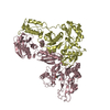
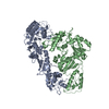
| Title | Crystal structure of HIV-1 RT bound to A 6-vinylpyrimidine inhibitor | ||||||
 Components Components |
| ||||||
 Keywords Keywords | TRANSFERASE / HYDROLASE / HIV-1 / inhibitor / RT | ||||||
| Function / homology |  Function and homology information Function and homology informationHIV-1 retropepsin / symbiont-mediated activation of host apoptosis / retroviral ribonuclease H / exoribonuclease H / exoribonuclease H activity / DNA integration / viral genome integration into host DNA / establishment of integrated proviral latency / RNA-directed DNA polymerase / RNA stem-loop binding ...HIV-1 retropepsin / symbiont-mediated activation of host apoptosis / retroviral ribonuclease H / exoribonuclease H / exoribonuclease H activity / DNA integration / viral genome integration into host DNA / establishment of integrated proviral latency / RNA-directed DNA polymerase / RNA stem-loop binding / viral penetration into host nucleus / host multivesicular body / RNA-directed DNA polymerase activity / RNA-DNA hybrid ribonuclease activity / Transferases; Transferring phosphorus-containing groups; Nucleotidyltransferases / host cell / viral nucleocapsid / DNA recombination / DNA-directed DNA polymerase / aspartic-type endopeptidase activity / Hydrolases; Acting on ester bonds / DNA-directed DNA polymerase activity / symbiont-mediated suppression of host gene expression / viral translational frameshifting / symbiont entry into host cell / lipid binding / host cell nucleus / host cell plasma membrane / virion membrane / structural molecule activity / proteolysis / DNA binding / zinc ion binding Similarity search - Function | ||||||
| Biological species |   Human immunodeficiency virus Human immunodeficiency virus | ||||||
| Method |  X-RAY DIFFRACTION / SYNCHROTRON / MOLECULAR REPLACEMENT / Resolution: 2.5 Å X-RAY DIFFRACTION / SYNCHROTRON / MOLECULAR REPLACEMENT / Resolution: 2.5 Å | ||||||
 Authors Authors | Ennifar, E. / Freisz, S. / Bec, G. / Dumas, P. / Botta, M. / Radi, M. | ||||||
 Citation Citation | Journal: Angew.Chem.Int.Ed.Engl. / Year: 2010 Title: Crystal Structure of HIV-1 Reverse Transcriptase Bound to a Non-Nucleoside Inhibitor with a Novel Mechanism of Action Authors: Freisz, S. / Bec, G. / Radi, M. / Wolff, P. / Crespan, E. / Angeli, L. / Dumas, P. / Maga, G. / Botta, M. / Ennifar, E. | ||||||
| History |
|
- Structure visualization
| Structure viewer | Molecule: MolmilJmol/JSmol |
|---|
- Downloads & links
-Download
| PDBx/mmCIF format | 3isn.cif.gz | 211.3 KB | Display | PDBx/mmCIF format |
|---|---|---|---|---|
| PDB format | pdb3isn.ent.gz | 169.1 KB | Display | PDB format |
| PDBx/mmJSON format | 3isn.json.gz | Tree view | PDBx/mmJSON format | |
| Others |  Other downloads Other downloads |
-Validation report
| Arichive directory | https://data.pdbj.org/pub/pdb/validation_reports/is/3isnftp://data.pdbj.org/pub/pdb/validation_reports/is/3isn | HTTPS FTP |
|---|
-Related structure data
| Related structure data | 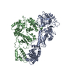 3ithC 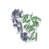 1dloS C: citing same article ( S: Starting model for refinement |
|---|---|
| Similar structure data |


-Links
 PDBj
PDBj


- Assembly
| Deposited unit | 
| ||||||||
|---|---|---|---|---|---|---|---|---|---|
| 1 |
| ||||||||
| Unit cell |
| ||||||||
| Components on special symmetry positions |
|
-Components
| #1: Protein | Mass: 64516.043 Da / Num. of mol.: 1 Source method: isolated from a genetically manipulated source Source: (gene. exp.) Human immunodeficiency virus / Strain: type 1 BH10 / Gene: gag-pol / Production host:  References: UniProt: P03366, RNA-directed DNA polymerase, DNA-directed DNA polymerase, ribonuclease H |
|---|---|
| #2: Protein | Mass: 49927.422 Da / Num. of mol.: 1 Source method: isolated from a genetically manipulated source Source: (gene. exp.) Human immunodeficiency virus / Strain: type 1 BH10 / Production host: |
| #3: Chemical | ChemComp-EDM /   Mass: 227.283 Da / Num. of mol.: 1 / Source method: obtained synthetically / Formula: C9H13N3O2S Mass: 227.283 Da / Num. of mol.: 1 / Source method: obtained synthetically / Formula: C9H13N3O2S |
| #4: Water | ChemComp-HOH /  Mass: 18.015 Da / Num. of mol.: 204 / Source method: isolated from a natural source / Formula: H2O Mass: 18.015 Da / Num. of mol.: 204 / Source method: isolated from a natural source / Formula: H2O |
| Has protein modification | N |
| Sequence details | E465Q IS MUTAGENESI |
-Experimental details
-Experiment
| Experiment | Method: X-RAY DIFFRACTION / Number of used crystals: 1 |
|---|
- Sample preparation
| Crystal | Density Matthews: 3.14 Å3/Da / Density % sol: 60.82 % |
|---|---|
| Crystal grow | Temperature: 277 K / Method: vapor diffusion, sitting drop / pH: 6.5 Details: 100mM Bis-Tris , 25% PEG 3350, 200mM ammonium sulfate, pH 6.5, VAPOR DIFFUSION, SITTING DROP, temperature 277K |
-Data collection
| Diffraction | Mean temperature: 90 K |
|---|---|
| Diffraction source | Source: SYNCHROTRON / Site: SLS  / Beamline: X06DA / Wavelength: 1.2824 Å / Beamline: X06DA / Wavelength: 1.2824 Å |
| Detector | Type: MARMOSAIC 225 mm CCD / Detector: CCD / Date: Dec 20, 2008 / Details: crystal |
| Radiation | Monochromator: crystal / Protocol: SINGLE WAVELENGTH / Monochromatic (M) / Laue (L): M / Scattering type: x-ray |
| Radiation wavelength | Wavelength: 1.2824 Å / Relative weight: 1 |
| Reflection | Resolution: 2.5→50 Å / Num. obs: 49675 / % possible obs: 82.3 % / Observed criterion σ(F): 0 / Observed criterion σ(I): 0 / Redundancy: 8.1 % / Biso Wilson estimate: 52.08 Å2 / Rmerge(I) obs: 0.076 / Rsym value: 0.062 / Net I/σ(I): 26.49 |
| Reflection shell | Resolution: 2.5→2.56 Å / Rmerge(I) obs: 0.542 / Mean I/σ(I) obs: 3.53 / Num. unique all: 40894 / Rsym value: 0.737 / % possible all: 67.4 |
- Processing
| Software |
| ||||||||||||||||||||||||||||||||||||||||||||||||||||||||||||||||||||||||||||||||||||||||||
|---|---|---|---|---|---|---|---|---|---|---|---|---|---|---|---|---|---|---|---|---|---|---|---|---|---|---|---|---|---|---|---|---|---|---|---|---|---|---|---|---|---|---|---|---|---|---|---|---|---|---|---|---|---|---|---|---|---|---|---|---|---|---|---|---|---|---|---|---|---|---|---|---|---|---|---|---|---|---|---|---|---|---|---|---|---|---|---|---|---|---|---|
| Refinement | Method to determine structure: MOLECULAR REPLACEMENT Starting model: PDB ID 1DLO Resolution: 2.5→49.102 Å / FOM work R set: 0.704 / SU ML: 0.41 / σ(F): 0 / σ(I): 0 / Phase error: 35.05 / Stereochemistry target values: ML
| ||||||||||||||||||||||||||||||||||||||||||||||||||||||||||||||||||||||||||||||||||||||||||
| Solvent computation | Shrinkage radii: 0.9 Å / VDW probe radii: 1.11 Å / Solvent model: FLAT BULK SOLVENT MODEL / Bsol: 50 Å2 / ksol: 0.324 e/Å3 | ||||||||||||||||||||||||||||||||||||||||||||||||||||||||||||||||||||||||||||||||||||||||||
| Displacement parameters | Biso max: 154.84 Å2 / Biso mean: 63.51 Å2 / Biso min: 25.42 Å2
| ||||||||||||||||||||||||||||||||||||||||||||||||||||||||||||||||||||||||||||||||||||||||||
| Refinement step | Cycle: LAST / Resolution: 2.5→49.102 Å
| ||||||||||||||||||||||||||||||||||||||||||||||||||||||||||||||||||||||||||||||||||||||||||
| Refine LS restraints |
| ||||||||||||||||||||||||||||||||||||||||||||||||||||||||||||||||||||||||||||||||||||||||||
| LS refinement shell | Refine-ID: X-RAY DIFFRACTION / Total num. of bins used: 14
|
