 Movie
Movie Controller


+ Open data

- Basic information
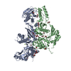
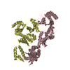
| Entry | Database: PDB / ID: 1hqe | ||||||
|---|---|---|---|---|---|---|---|
| Title | HUMAN IMMUNODEFICIENCY VIRUS TYPE 1 | ||||||
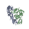
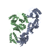
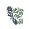
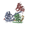
 Components Components | (POL POLYPROTEIN) x 2 | ||||||
 Keywords Keywords | TRANSFERASE / NUCLEOTIDYLTRANSFERASE | ||||||
| Function / homology |  Function and homology information Function and homology informationHIV-1 retropepsin / symbiont-mediated activation of host apoptosis / retroviral ribonuclease H / exoribonuclease H / exoribonuclease H activity / DNA integration / viral genome integration into host DNA / establishment of integrated proviral latency / RNA-directed DNA polymerase / RNA stem-loop binding ...HIV-1 retropepsin / symbiont-mediated activation of host apoptosis / retroviral ribonuclease H / exoribonuclease H / exoribonuclease H activity / DNA integration / viral genome integration into host DNA / establishment of integrated proviral latency / RNA-directed DNA polymerase / RNA stem-loop binding / viral penetration into host nucleus / host multivesicular body / RNA-directed DNA polymerase activity / RNA-DNA hybrid ribonuclease activity / Transferases; Transferring phosphorus-containing groups; Nucleotidyltransferases / host cell / viral nucleocapsid / DNA recombination / DNA-directed DNA polymerase / aspartic-type endopeptidase activity / Hydrolases; Acting on ester bonds / DNA-directed DNA polymerase activity / symbiont-mediated suppression of host gene expression / viral translational frameshifting / symbiont entry into host cell / lipid binding / host cell nucleus / host cell plasma membrane / virion membrane / structural molecule activity / proteolysis / DNA binding / zinc ion binding Similarity search - Function | ||||||
| Biological species |   Human immunodeficiency virus 1 Human immunodeficiency virus 1 | ||||||
| Method |  X-RAY DIFFRACTION / SYNCHROTRON / MOLECULAR REPLACEMENT / Resolution: 2.7 Å X-RAY DIFFRACTION / SYNCHROTRON / MOLECULAR REPLACEMENT / Resolution: 2.7 Å | ||||||
 Authors Authors | Ding, J. / Hsiou, Y. / Arnold, E. | ||||||
 Citation Citation | Journal: J.Mol.Biol. / Year: 2001 Title: The Lys103Asn mutation of HIV-1 RT: a novel mechanism of drug resistance. Authors: Hsiou, Y. / Ding, J. / Das, K. / Clark Jr., A.D. / Boyer, P.L. / Lewi, P. / Janssen, P.A. / Kleim, J.P. / Rosner, M. / Hughes, S.H. / Arnold, E. #1: Journal: Structure / Year: 1996Title: Structure of Unliganded HIV-1 Reverse Transcriptase at 2.7 A Resolution: Implications of Conformational Changes for Polymerization and Inhibition Mechanisms Authors: Hsiou, Y. / Ding, J. / Das, K. / Clark Jr., A.D. / Hughes, S.H. / Arnold, E. | ||||||
| History |
|
- Structure visualization
| Structure viewer | Molecule: MolmilJmol/JSmol |
|---|
- Downloads & links
-Download
| PDBx/mmCIF format | 1hqe.cif.gz | 204.7 KB | Display | PDBx/mmCIF format |
|---|---|---|---|---|
| PDB format | pdb1hqe.ent.gz | 163 KB | Display | PDB format |
| PDBx/mmJSON format | 1hqe.json.gz | Tree view | PDBx/mmJSON format | |
| Others |  Other downloads Other downloads |
-Validation report
| Arichive directory | https://data.pdbj.org/pub/pdb/validation_reports/hq/1hqeftp://data.pdbj.org/pub/pdb/validation_reports/hq/1hqe | HTTPS FTP |
|---|
-Related structure data
| Related structure data | 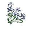 1hpzC 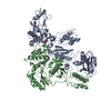 1hquC 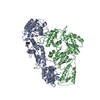 1dloS C: citing same article ( S: Starting model for refinement |
|---|---|
| Similar structure data |
-Links
 PDBj
PDBj


- Assembly
| Deposited unit | 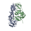
| ||||||||
|---|---|---|---|---|---|---|---|---|---|
| 1 |
| ||||||||
| Unit cell |
|
-Components
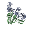
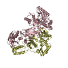
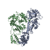
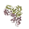
| #1: Protein | Mass: 64485.887 Da / Num. of mol.: 1 / Fragment: P66 SUBUNIT / Mutation: K103N, C280S Source method: isolated from a genetically manipulated source Source: (gene. exp.) Human immunodeficiency virus 1 / Genus: Lentivirus / Description: HIV-1 CLONE 12 / Production host:  |
|---|---|
| #2: Protein | Mass: 50266.684 Da / Num. of mol.: 1 / Fragment: P51 SUBUNIT / Mutation: K103N, C280S Source method: isolated from a genetically manipulated source Source: (gene. exp.) Human immunodeficiency virus 1 / Genus: Lentivirus / Description: HIV-1 CLONE 12 / Production host: |
| #3: Water | ChemComp-HOH /  Mass: 18.015 Da / Num. of mol.: 182 / Source method: isolated from a natural source / Formula: H2O Mass: 18.015 Da / Num. of mol.: 182 / Source method: isolated from a natural source / Formula: H2O |
-Experimental details
-Experiment
| Experiment | Method: X-RAY DIFFRACTION / Number of used crystals: 1 |
|---|
- Sample preparation
| Crystal | Density Matthews: 3.25 Å3/Da / Density % sol: 62.1 % | ||||||||||||||||||||||||||||||||||||||||||||||||||||||||||||||||||
|---|---|---|---|---|---|---|---|---|---|---|---|---|---|---|---|---|---|---|---|---|---|---|---|---|---|---|---|---|---|---|---|---|---|---|---|---|---|---|---|---|---|---|---|---|---|---|---|---|---|---|---|---|---|---|---|---|---|---|---|---|---|---|---|---|---|---|---|
| Crystal grow | Temperature: 277 K / Method: vapor diffusion, hanging drop / pH: 6.8 Details: Bis-Tris.propane, ammoniun sulfate, glycerol, PEG 8000, pH 6.8, VAPOR DIFFUSION, HANGING DROP, temperature 277K | ||||||||||||||||||||||||||||||||||||||||||||||||||||||||||||||||||
| Crystal grow | *PLUS pH: 8 / Details: used microseeding | ||||||||||||||||||||||||||||||||||||||||||||||||||||||||||||||||||
| Components of the solutions | *PLUS
|
-Data collection
| Diffraction | Mean temperature: 108 K |
|---|---|
| Diffraction source | Source: SYNCHROTRON / Site: CHESS  / Beamline: F1 / Wavelength: 0.91 Å / Beamline: F1 / Wavelength: 0.91 Å |
| Detector | Type: ADSC QUANTUM 4 / Detector: CCD / Date: Jan 28, 1997 |
| Radiation | Protocol: SINGLE WAVELENGTH / Monochromatic (M) / Laue (L): M / Scattering type: x-ray |
| Radiation wavelength | Wavelength: 0.91 Å / Relative weight: 1 |
| Reflection | Resolution: 2.7→50 Å / Num. obs: 37652 / % possible obs: 92.2 % / Observed criterion σ(F): 0 / Observed criterion σ(I): 0 / Redundancy: 8.6 % / Rmerge(I) obs: 0.101 / Net I/σ(I): 6.6 |
| Reflection | *PLUS Lowest resolution: 50 Å |
| Reflection shell | *PLUS Highest resolution: 2.7 Å / Lowest resolution: 2.8 Å / % possible obs: 87.6 % / Rmerge(I) obs: 0.376 |
- Processing
| Software |
| |||||||||||||||||||||
|---|---|---|---|---|---|---|---|---|---|---|---|---|---|---|---|---|---|---|---|---|---|---|
| Refinement | Method to determine structure: MOLECULAR REPLACEMENT Starting model: PDB entry 1DLO Resolution: 2.7→25 Å / Isotropic thermal model: isotropic / σ(F): 2 / Stereochemistry target values: Engh and Huber
| |||||||||||||||||||||
| Displacement parameters | Biso mean: 64.8 Å2 | |||||||||||||||||||||
| Refinement step | Cycle: LAST / Resolution: 2.7→25 Å
| |||||||||||||||||||||
| Refine LS restraints |
| |||||||||||||||||||||
| Software | *PLUS Name: X-PLOR / Version: 3.843 / Classification: refinement | |||||||||||||||||||||
| Refinement | *PLUS Highest resolution: 2.7 Å / Lowest resolution: 25 Å / σ(F): 2 / % reflection Rfree: 3.5 % / Rfactor obs: 0.25 / Rfactor Rwork: 0.25 | |||||||||||||||||||||
| Solvent computation | *PLUS | |||||||||||||||||||||
| Displacement parameters | *PLUS Biso mean: 64.8 Å2 | |||||||||||||||||||||
| Refine LS restraints | *PLUS
| |||||||||||||||||||||
| LS refinement shell | *PLUS Highest resolution: 2.7 Å / Lowest resolution: 2.8 Å / Rfactor Rfree: 0.385 / Rfactor obs: 0.359 |
