 Movie
Movie Controller

[English] 日本語
 Yorodumi
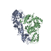
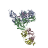
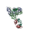
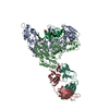
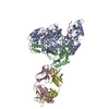
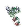
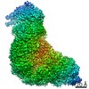
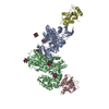
- PDB-1j5o: CRYSTAL STRUCTURE OF MET184ILE MUTANT OF HIV-1 REVERSE TRANSCRIPT... -
+ Open data

- Basic information
| Entry | Database: PDB / ID: 1j5o | |||||||||
|---|---|---|---|---|---|---|---|---|---|---|
| Title | CRYSTAL STRUCTURE OF MET184ILE MUTANT OF HIV-1 REVERSE TRANSCRIPTASE IN COMPLEX WITH DOUBLE STRANDED DNA TEMPLATE-PRIMER | |||||||||
 Components Components |
| |||||||||
 Keywords Keywords | TRANSFERASE/IMMUNE SYSTEM/DNA / HIV / REVERSE TRANSCRIPTASE / MET184ILE / 3TC / PROTEIN-DNA COMPLEX / DRUG RESISTANCE / M184I / TRANSFERASE-IMMUNE SYSTEM-DNA COMPLEX | |||||||||
| Function / homology |  Function and homology information Function and homology informationHIV-1 retropepsin / symbiont-mediated activation of host apoptosis / retroviral ribonuclease H / exoribonuclease H / exoribonuclease H activity / DNA integration / viral genome integration into host DNA / establishment of integrated proviral latency / RNA-directed DNA polymerase / RNA stem-loop binding ...HIV-1 retropepsin / symbiont-mediated activation of host apoptosis / retroviral ribonuclease H / exoribonuclease H / exoribonuclease H activity / DNA integration / viral genome integration into host DNA / establishment of integrated proviral latency / RNA-directed DNA polymerase / RNA stem-loop binding / viral penetration into host nucleus / host multivesicular body / RNA-directed DNA polymerase activity / RNA-DNA hybrid ribonuclease activity / Transferases; Transferring phosphorus-containing groups; Nucleotidyltransferases / host cell / viral nucleocapsid / DNA recombination / DNA-directed DNA polymerase / aspartic-type endopeptidase activity / Hydrolases; Acting on ester bonds / DNA-directed DNA polymerase activity / symbiont-mediated suppression of host gene expression / viral translational frameshifting / symbiont entry into host cell / lipid binding / host cell nucleus / host cell plasma membrane / virion membrane / structural molecule activity / proteolysis / DNA binding / zinc ion binding Similarity search - Function | |||||||||
| Biological species |   Human immunodeficiency virus 1 Human immunodeficiency virus 1 | |||||||||
| Method |  X-RAY DIFFRACTION / SYNCHROTRON / Resolution: 3.5 Å X-RAY DIFFRACTION / SYNCHROTRON / Resolution: 3.5 Å | |||||||||
 Authors Authors | Sarafianos, S.G. / Das, K. / Arnold, E. | |||||||||
 Citation Citation | Journal: Proc.Natl.Acad.Sci.USA / Year: 1999 Title: Lamivudine (3TC) resistance in HIV-1 reverse transcriptase involves steric hindrance with beta-branched amino acids. Authors: Sarafianos, S.G. / Das, K. / Clark Jr., A.D. / Ding, J. / Boyer, P.L. / Hughes, S.H. / Arnold, E. #1: Journal: J.Mol.Biol. / Year: 1998Title: Structure and Functional Implications of the Polymerase Active Site Region in a Complex of HIV-1 RT with a Double-Stranded DNA template-primer and an Antibody Fab Fragment at 2.8 Angstroms Resolution Authors: Ding, J. / Das, K. / Hsiou, Y. / Sarafianos, S.G. / Clark Jr., A.D. / Jacobo-Molina, A. / Tantillo, C. / Hughes, S.H. / Arnold, E. #2: Journal: Chem.Biol. / Year: 1999Title: Touching the Heart of HIV-1 Drug Resistance: The Fingers Close Down on the dNTP at the Polymerase Active Site Authors: Sarafianos, S.G. / Das, K. / Ding, J. / Boyer, P.L. / Hughes, S.H. / Arnold, E. | |||||||||
| History |
|
- Structure visualization
| Structure viewer | Molecule: MolmilJmol/JSmol |
|---|
- Downloads & links
-Download
| PDBx/mmCIF format | 1j5o.cif.gz | 306.9 KB | Display | PDBx/mmCIF format |
|---|---|---|---|---|
| PDB format | pdb1j5o.ent.gz | 239.7 KB | Display | PDB format |
| PDBx/mmJSON format | 1j5o.json.gz | Tree view | PDBx/mmJSON format | |
| Others |  Other downloads Other downloads |
-Validation report
| Arichive directory | https://data.pdbj.org/pub/pdb/validation_reports/j5/1j5oftp://data.pdbj.org/pub/pdb/validation_reports/j5/1j5o | HTTPS FTP |
|---|
-Related structure data


-Links
 PDBj
PDBj


- Assembly
| Deposited unit | 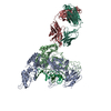
| ||||||||
|---|---|---|---|---|---|---|---|---|---|
| 1 |
| ||||||||
| Unit cell |
|
-Components
-DNA chain , 2 types, 2 molecules TP
| #1: DNA chain | Mass: 5864.801 Da / Num. of mol.: 1 / Source method: obtained synthetically |
|---|---|
| #2: DNA chain | Mass: 5484.528 Da / Num. of mol.: 1 / Source method: obtained synthetically |
-Protein , 2 types, 2 molecules AB
| #3: Protein | Mass: 64256.613 Da / Num. of mol.: 1 / Mutation: M184I,C280S Source method: isolated from a genetically manipulated source Source: (gene. exp.) Human immunodeficiency virus 1 / Genus: Lentivirus / Production host:  |
|---|---|
| #4: Protein | Mass: 50263.723 Da / Num. of mol.: 1 / Mutation: M184I,C280S Source method: isolated from a genetically manipulated source Source: (gene. exp.) Human immunodeficiency virus 1 / Genus: Lentivirus / Production host: |
-Antibody , 2 types, 2 molecules LH
| #5: Antibody | Mass: 22984.068 Da / Num. of mol.: 1 / Fragment: FAB28 / Source method: isolated from a natural source / Source: (natural) |
|---|---|
| #6: Antibody | Mass: 23457.156 Da / Num. of mol.: 1 / Fragment: FAB28 / Source method: isolated from a natural source / Source: (natural) |
-Details
| Has protein modification | Y |
|---|
-Experimental details
-Experiment
| Experiment | Method: X-RAY DIFFRACTION / Number of used crystals: 10 |
|---|
- Sample preparation
| Crystal | Density Matthews: 5.32 Å3/Da / Density % sol: 76.88 % | ||||||||||||||||||||||||||||||
|---|---|---|---|---|---|---|---|---|---|---|---|---|---|---|---|---|---|---|---|---|---|---|---|---|---|---|---|---|---|---|---|
| Crystal grow | Temperature: 277 K / Method: vapor diffusion, hanging drop / pH: 5.6 Details: 30 % SATURATED AMMONIUM SULFATE, 100 MM CACODYLATE PH 5.6, VAPOR DIFFUSION, HANGING DROP, temperature 277K | ||||||||||||||||||||||||||||||
| Crystal grow | *PLUS Temperature: 4 ℃ | ||||||||||||||||||||||||||||||
| Components of the solutions | *PLUS
|
-Data collection
| Diffraction |
| |||||||||||||||
|---|---|---|---|---|---|---|---|---|---|---|---|---|---|---|---|---|
| Diffraction source |
| |||||||||||||||
| Detector |
| |||||||||||||||
| Radiation |
| |||||||||||||||
| Radiation wavelength |
| |||||||||||||||
| Reflection | Resolution: 3.5→40 Å / Num. all: 46038 / Num. obs: 42062 / % possible obs: 91.4 % / Observed criterion σ(F): 0 / Observed criterion σ(I): 0 / Redundancy: 5.91 % / Rmerge(I) obs: 0.118 / Net I/σ(I): 11.2 | |||||||||||||||
| Reflection shell | Resolution: 3.5→3.63 Å / Redundancy: 2.8 % / Rmerge(I) obs: 0.4 / % possible all: 83 | |||||||||||||||
| Reflection | *PLUS Lowest resolution: 40 Å / % possible obs: 91.5 % | |||||||||||||||
| Reflection shell | *PLUS % possible obs: 83 % |

- Processing
| Software |
| ||||||||||||||||||||||||||||||||||||||||||||||||||||||||||||
|---|---|---|---|---|---|---|---|---|---|---|---|---|---|---|---|---|---|---|---|---|---|---|---|---|---|---|---|---|---|---|---|---|---|---|---|---|---|---|---|---|---|---|---|---|---|---|---|---|---|---|---|---|---|---|---|---|---|---|---|---|---|
| Refinement | Resolution: 3.5→10 Å / σ(F): 1 / σ(I): 1
| ||||||||||||||||||||||||||||||||||||||||||||||||||||||||||||
| Refinement step | Cycle: LAST / Resolution: 3.5→10 Å
| ||||||||||||||||||||||||||||||||||||||||||||||||||||||||||||
| Refine LS restraints |
| ||||||||||||||||||||||||||||||||||||||||||||||||||||||||||||
| Refinement | *PLUS Lowest resolution: 10 Å | ||||||||||||||||||||||||||||||||||||||||||||||||||||||||||||
| Solvent computation | *PLUS | ||||||||||||||||||||||||||||||||||||||||||||||||||||||||||||
| Displacement parameters | *PLUS |
