 Movie
Movie Controller


+ Open data

- Basic information
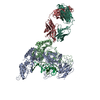
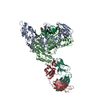
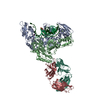
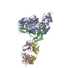
| Entry | Database: PDB / ID: 2hmi | |||||||||
|---|---|---|---|---|---|---|---|---|---|---|
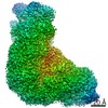
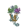
| Title | HIV-1 REVERSE TRANSCRIPTASE/FRAGMENT OF FAB 28/DNA COMPLEX | |||||||||
 Components Components |
| |||||||||
 Keywords Keywords | IMMUNE SYSTEM/DNA / AIDS / HIV-1 / RT / POLYMERASE / IMMUNE SYSTEM-DNA COMPLEX | |||||||||
| Function / homology |  Function and homology information Function and homology informationHIV-1 retropepsin / symbiont-mediated activation of host apoptosis / retroviral ribonuclease H / exoribonuclease H / exoribonuclease H activity / DNA integration / viral genome integration into host DNA / establishment of integrated proviral latency / RNA-directed DNA polymerase / RNA stem-loop binding ...HIV-1 retropepsin / symbiont-mediated activation of host apoptosis / retroviral ribonuclease H / exoribonuclease H / exoribonuclease H activity / DNA integration / viral genome integration into host DNA / establishment of integrated proviral latency / RNA-directed DNA polymerase / RNA stem-loop binding / viral penetration into host nucleus / host multivesicular body / RNA-directed DNA polymerase activity / RNA-DNA hybrid ribonuclease activity / Transferases; Transferring phosphorus-containing groups; Nucleotidyltransferases / host cell / viral nucleocapsid / DNA recombination / DNA-directed DNA polymerase / aspartic-type endopeptidase activity / Hydrolases; Acting on ester bonds / DNA-directed DNA polymerase activity / symbiont-mediated suppression of host gene expression / viral translational frameshifting / symbiont entry into host cell / lipid binding / host cell nucleus / host cell plasma membrane / virion membrane / structural molecule activity / proteolysis / DNA binding / zinc ion binding Similarity search - Function | |||||||||
| Biological species |   Human immunodeficiency virus 1 Human immunodeficiency virus 1 | |||||||||
| Method |  X-RAY DIFFRACTION / SYNCHROTRON / Resolution: 2.8 Å X-RAY DIFFRACTION / SYNCHROTRON / Resolution: 2.8 Å | |||||||||
 Authors Authors | Ding, J. / Arnold, E. | |||||||||
 Citation Citation | Journal: J.Mol.Biol. / Year: 1998 Title: Structure and functional implications of the polymerase active site region in a complex of HIV-1 RT with a double-stranded DNA template-primer and an antibody Fab fragment at 2.8 A resolution. Authors: Ding, J. / Das, K. / Hsiou, Y. / Sarafianos, S.G. / Clark Jr., A.D. / Jacobo-Molina, A. / Tantillo, C. / Hughes, S.H. / Arnold, E. #1: Journal: Structure / Year: 1996Title: Structure of Unliganded HIV-1 Reverse Transcriptase at 2.7 A Resolution: Implications of Conformational Changes for Polymerization and Inhibition Mechanisms Authors: Hsiou, Y. / Ding, J. / Das, K. / Clark Junior, A.D. / Hughes, S.H. / Arnold, E. #2: Journal: Methods Enzymol. / Year: 1995Title: Crystallization of Human Immunodeficiency Virus Type 1 Reverse Transcriptase with and without Nucleic Acid Substrates, Inhibitors, and an Antibody Fab Fragment Authors: Clark Junior, A.D. / Jacobo-Molina, A. / Clark, P. / Hughes, S.H. / Arnold, E. #3: Journal: Structure / Year: 1995Title: Structure of HIV-1 Reverse Transcriptase in a Complex with the Non-Nucleoside Inhibitor Alpha-Apa R 95845 at 2.8 A Resolution Authors: Ding, J. / Das, K. / Tantillo, C. / Zhang, W. / Clark Junior, A.D. / Jessen, S. / Lu, X. / Hsiou, Y. / Jacobo-Molina, A. / Andries, K. / Pauwels, R. / Moereels, H. / Koymans, L. / Janssen, P. ...Authors: Ding, J. / Das, K. / Tantillo, C. / Zhang, W. / Clark Junior, A.D. / Jessen, S. / Lu, X. / Hsiou, Y. / Jacobo-Molina, A. / Andries, K. / Pauwels, R. / Moereels, H. / Koymans, L. / Janssen, P.A.J. / Smith Junior, R.H. / Koepke, M.K. / Michejda, C.J. / Hughes, S.H. / Arnold, E. #4: Journal: Nat.Struct.Biol. / Year: 1995Title: Structure of HIV-1 RT/TIBO R 86183 Complex Reveals Similarity in the Binding of Diverse Nonnucleoside Inhibitors Authors: Ding, J. / Das, K. / Moereels, H. / Koymans, L. / Andries, K. / Janssen, P.A. / Hughes, S.H. / Arnold, E. #5: Journal: Proc.Natl.Acad.Sci.USA / Year: 1993Title: Crystal Structure of Human Immunodeficiency Virus Type 1 Reverse Transcriptase Complexed with Double-Stranded DNA at 3.0 A Resolution Shows Bent DNA Authors: Jacobo-Molina, A. / Ding, J. / Nanni, R.G. / Clark Junior, A.D. / Lu, X. / Tantillo, C. / Williams, R.L. / Kamer, G. / Ferris, A.L. / Clark, P. / Hizi, A. / Hughes, S.H. / Arnold, E. #6: Journal: Nature / Year: 1992Title: Structure of HIV-1 Reverse Transcriptase/DNA Complex at 7 A Resolution Showing Active Site Locations Authors: Arnold, E. / Jacobo-Molina, A. / Nanni, R.G. / Williams, R.L. / Lu, X. / Ding, J. / Clark Junior, A.D. / Zhang, A. / Ferris, A.L. / Clark, P. / Hizi, A. / Hughes, S.H. | |||||||||
| History |
|
- Structure visualization
| Structure viewer | Molecule: MolmilJmol/JSmol |
|---|
- Downloads & links
-Download
| PDBx/mmCIF format | 2hmi.cif.gz | 304.3 KB | Display | PDBx/mmCIF format |
|---|---|---|---|---|
| PDB format | pdb2hmi.ent.gz | 238.3 KB | Display | PDB format |
| PDBx/mmJSON format | 2hmi.json.gz | Tree view | PDBx/mmJSON format | |
| Others |  Other downloads Other downloads |
-Validation report
| Arichive directory | https://data.pdbj.org/pub/pdb/validation_reports/hm/2hmiftp://data.pdbj.org/pub/pdb/validation_reports/hm/2hmi | HTTPS FTP |
|---|
-Related structure data
| Similar structure data |
|---|


-Links
 PDBj
PDBj


- Assembly
| Deposited unit | 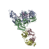
| ||||||||||
|---|---|---|---|---|---|---|---|---|---|---|---|
| 1 |
| ||||||||||
| Unit cell |
|
-Components
-DNA chain , 2 types, 2 molecules EF
| #1: DNA chain | Mass: 5864.801 Da / Num. of mol.: 1 / Source method: obtained synthetically |
|---|---|
| #2: DNA chain | Mass: 5484.528 Da / Num. of mol.: 1 / Source method: obtained synthetically |
-Protein , 2 types, 2 molecules AB
| #3: Protein | Mass: 64274.652 Da / Num. of mol.: 1 / Mutation: C280S Source method: isolated from a genetically manipulated source Source: (gene. exp.) Human immunodeficiency virus 1 / Genus: Lentivirus / Strain: BH10 ISOLATE / Production host:  |
|---|---|
| #4: Protein | Mass: 50281.762 Da / Num. of mol.: 1 / Mutation: C280S Source method: isolated from a genetically manipulated source Source: (gene. exp.) Human immunodeficiency virus 1 / Genus: Lentivirus / Strain: BH10 ISOLATE / Production host: |
-Antibody , 2 types, 2 molecules CD
| #5: Antibody | Mass: 22984.068 Da / Num. of mol.: 1 / Fragment: FAB FRAGMENT / Source method: isolated from a natural source / Source: (natural) |
|---|---|
| #6: Antibody | Mass: 23457.156 Da / Num. of mol.: 1 / Fragment: FAB FRAGMENT / Source method: isolated from a natural source / Source: (natural) |
-Details
| Compound details | HIV-1 RT IS COMPOSED OF TWO SUBUNITS OF 66 KDA AND 51 KDA, DESIGNATED AS P66 (CHAIN A) AND P51 ...HIV-1 RT IS COMPOSED OF TWO SUBUNITS OF 66 KDA AND 51 KDA, DESIGNATED |
|---|---|
| Has protein modification | Y |
| Sequence details | THE BOUND DOUBLE-STRANDED DNA IS A 19-MER/18-MER TEMPLATE- PRIMER. BOTH THE TEMPLATE STRAND AND THE ...THE BOUND DOUBLE-STRANDED DNA IS A 19-MER/18-MER TEMPLATE- PRIMER. BOTH THE TEMPLATE STRAND AND THE PRIMER STRAND ARE NUMBERED IN THE 5'-3' DIRECTION. THE FIRST NUCLEOTIDE |
-Experimental details
-Experiment
| Experiment | Method: X-RAY DIFFRACTION / Number of used crystals: 1 |
|---|
- Sample preparation
| Crystal | Density Matthews: 5.29 Å3/Da / Density % sol: 75.6 % Description: INTENSITY-INTEGRATION SOFTWARE USED WAS A MODIFIED VERSION OF THE PURDUE OSCILLATION FILM PROCESSING PACKAGE AND DENZO | ||||||||||||||||||||||||
|---|---|---|---|---|---|---|---|---|---|---|---|---|---|---|---|---|---|---|---|---|---|---|---|---|---|
| Crystal | *PLUS | ||||||||||||||||||||||||
| Crystal grow | *PLUS Method: vapor diffusion, hanging drop / Details: Jacobo-Molina, A., (1991) Biochemistry, 30, 6351. / pH: 8.8 | ||||||||||||||||||||||||
| Components of the solutions | *PLUS
|
-Data collection
| Diffraction source | Source: SYNCHROTRON / Site: CHESS  / Beamline: F1 / Beamline: F1 |
|---|---|
| Detector | Type: FUJI / Detector: IMAGE PLATE / Date: 1992 |
| Radiation | Protocol: SINGLE WAVELENGTH / Monochromatic (M) / Laue (L): M / Scattering type: x-ray |
| Radiation wavelength | Relative weight: 1 |
| Reflection | Redundancy: 4.3 % / Biso Wilson estimate: 21.8 Å2 / Rmerge(I) obs: 0.13 |
| Reflection | *PLUS Highest resolution: 2.8 Å / Num. obs: 76187 / % possible obs: 86.6 % / Observed criterion σ(I): 2 |
- Processing
| Software |
| ||||||||||||||||||||||||||||||||||||||||||||||||||||||||||||
|---|---|---|---|---|---|---|---|---|---|---|---|---|---|---|---|---|---|---|---|---|---|---|---|---|---|---|---|---|---|---|---|---|---|---|---|---|---|---|---|---|---|---|---|---|---|---|---|---|---|---|---|---|---|---|---|---|---|---|---|---|---|
| Refinement | Resolution: 2.8→8 Å / Rfactor Rfree error: 0.006 / Data cutoff high absF: 100000 / Data cutoff low absF: 125 / Isotropic thermal model: RESTRAINED / Cross valid method: THROUGHOUT / σ(F): 2
| ||||||||||||||||||||||||||||||||||||||||||||||||||||||||||||
| Displacement parameters | Biso mean: 36.9 Å2
| ||||||||||||||||||||||||||||||||||||||||||||||||||||||||||||
| Refine analyze |
| ||||||||||||||||||||||||||||||||||||||||||||||||||||||||||||
| Refinement step | Cycle: LAST / Resolution: 2.8→8 Å
| ||||||||||||||||||||||||||||||||||||||||||||||||||||||||||||
| Refine LS restraints |
| ||||||||||||||||||||||||||||||||||||||||||||||||||||||||||||
| LS refinement shell | Resolution: 2.8→2.9 Å / Rfactor Rfree error: 0.031 / Total num. of bins used: 10
| ||||||||||||||||||||||||||||||||||||||||||||||||||||||||||||
| Xplor file |
| ||||||||||||||||||||||||||||||||||||||||||||||||||||||||||||
| Software | *PLUS Name: X-PLOR / Version: 3.1 / Classification: refinement | ||||||||||||||||||||||||||||||||||||||||||||||||||||||||||||
| Refinement | *PLUS Rfactor obs: 0.271 | ||||||||||||||||||||||||||||||||||||||||||||||||||||||||||||
| Solvent computation | *PLUS | ||||||||||||||||||||||||||||||||||||||||||||||||||||||||||||
| Displacement parameters | *PLUS | ||||||||||||||||||||||||||||||||||||||||||||||||||||||||||||
| Refine LS restraints | *PLUS
|
