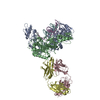
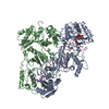
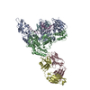
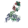
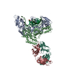
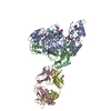
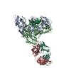
[English] 日本語
Yorodumi
- PDB-1n5y: HIV-1 Reverse Transcriptase Crosslinked to Post-Translocation AZT... -

+
Open data


ID or keywords:

Loading...

-
Basic information

Entry
Database: PDB / ID: 1n5y
TitleHIV-1 Reverse Transcriptase Crosslinked to Post-Translocation AZTMP-Terminated DNA (Complex P)
Components
  • (REVERSE TRANSCRIPTASE) x 2
  • (monoclonal antibody ...) x 2
  • 5'-D(*A*CP*AP*GP*TP*CP*CP*CP*TP*GP*TP*TP*CP*GP*GP*(MRG)P*CP*GP*CP*CP*(ATM))-3'
  • 5'-D(*AP*TP*GP*C*TP*AP*GP*GP*CP*GP*CP*CP*CP*GP*AP*AP*CP*AP*GP*GP*GP*AP*CP*TP*GP*TP*G)-3'
KeywordsTRANSFERASE/IMMUNE SYSTEM/DNA / HIV / translocation / nucleotide excision / drug resistance / complex P / TRANSFERASE-IMMUNE SYSTEM-DNA COMPLEX
Function / homology
Function and homology information


humoral immune response mediated by circulating immunoglobulin / phagocytosis, recognition / positive regulation of type IIa hypersensitivity / positive regulation of type I hypersensitivity / antibody-dependent cellular cytotoxicity / Fc-gamma receptor I complex binding / phagocytosis, engulfment / immunoglobulin complex, circulating / IgG immunoglobulin complex / immunoglobulin receptor binding ...humoral immune response mediated by circulating immunoglobulin / phagocytosis, recognition / positive regulation of type IIa hypersensitivity / positive regulation of type I hypersensitivity / antibody-dependent cellular cytotoxicity / Fc-gamma receptor I complex binding / phagocytosis, engulfment / immunoglobulin complex, circulating / IgG immunoglobulin complex / immunoglobulin receptor binding / immunoglobulin mediated immune response / positive regulation of phagocytosis / complement activation, classical pathway / antigen binding / B cell differentiation / HIV-1 retropepsin / : / retroviral ribonuclease H / exoribonuclease H / : / exoribonuclease H activity / host multivesicular body / DNA integration / RNA-directed DNA polymerase / viral genome integration into host DNA / viral penetration into host nucleus / establishment of integrated proviral latency / RNA-directed DNA polymerase activity / positive regulation of immune response / Transferases; Transferring phosphorus-containing groups; Nucleotidyltransferases / RNA-DNA hybrid ribonuclease activity / antibacterial humoral response / viral nucleocapsid / DNA recombination / Hydrolases; Acting on ester bonds / DNA-directed DNA polymerase / aspartic-type endopeptidase activity / DNA-directed DNA polymerase activity / defense response to bacterium / symbiont entry into host cell / symbiont-mediated suppression of host gene expression / external side of plasma membrane / lipid binding / host cell nucleus / structural molecule activity / host cell plasma membrane / virion membrane / proteolysis / DNA binding / extracellular space / RNA binding / zinc ion binding / extracellular region / membrane / plasma membrane / cytoplasm
Similarity search - Function
HIV Type 1 Reverse Transcriptase, subunit A, domain 1 / HIV Type 1 Reverse Transcriptase; Chain A, domain 1 / Reverse transcriptase/Diguanylate cyclase domain / Ribonuclease H-like superfamily/Ribonuclease H / Reverse transcriptase connection / Reverse transcriptase connection domain / Reverse transcriptase thumb / Reverse transcriptase thumb domain / Integrase Zinc binding domain / Zinc finger integrase-type profile. ...HIV Type 1 Reverse Transcriptase, subunit A, domain 1 / HIV Type 1 Reverse Transcriptase; Chain A, domain 1 / Reverse transcriptase/Diguanylate cyclase domain / Ribonuclease H-like superfamily/Ribonuclease H / Reverse transcriptase connection / Reverse transcriptase connection domain / Reverse transcriptase thumb / Reverse transcriptase thumb domain / Integrase Zinc binding domain / Zinc finger integrase-type profile. / Integrase-like, N-terminal / Integrase DNA binding domain / Integrase, C-terminal domain superfamily, retroviral / Integrase, N-terminal zinc-binding domain / Integrase, C-terminal, retroviral / Integrase DNA binding domain profile. / Immunodeficiency lentiviral matrix, N-terminal / gag gene protein p17 (matrix protein) / RNase H / Integrase core domain / Integrase, catalytic core / Integrase catalytic domain profile. / Retroviral nucleocapsid Gag protein p24, C-terminal domain / Gag protein p24 C-terminal domain / Retropepsin-like catalytic domain / Matrix protein, lentiviral and alpha-retroviral, N-terminal / Ribonuclease H domain / RNase H type-1 domain profile. / Reverse transcriptase (RNA-dependent DNA polymerase) / Reverse transcriptase domain / Reverse transcriptase (RT) catalytic domain profile. / Retropepsins / Retroviral aspartyl protease / Aspartyl protease, retroviral-type family profile. / Peptidase A2A, retrovirus, catalytic / Retrovirus capsid, C-terminal / Retroviral matrix protein / Retrovirus capsid, N-terminal / zinc finger / Zinc knuckle / Zinc finger, CCHC-type superfamily / Zinc finger, CCHC-type / Zinc finger CCHC-type profile. / Nucleotidyltransferase; domain 5 / Immunoglobulin/major histocompatibility complex, conserved site / Immunoglobulins and major histocompatibility complex proteins signature. / Immunoglobulin C-Type / Immunoglobulin C1-set / Immunoglobulin C1-set domain / Ribonuclease H superfamily / Aspartic peptidase, active site / Eukaryotic and viral aspartyl proteases active site. / Aspartic peptidase domain superfamily / Ribonuclease H-like superfamily / Reverse transcriptase/Diguanylate cyclase domain / Alpha-Beta Plaits / Ig-like domain profile. / Immunoglobulin-like domain / Immunoglobulin-like domain superfamily / Immunoglobulins / Roll / DNA/RNA polymerase superfamily / Immunoglobulin-like fold / Immunoglobulin-like / Sandwich / 2-Layer Sandwich / Mainly Beta / Alpha Beta
Similarity search - Domain/homology
DNA / DNA (> 10) / Immunoglobulin kappa constant / Ig gamma-1 chain C region secreted form / Gag-Pol polyprotein
Similarity search - Component
Biological speciesHuman immunodeficiency virus 1
Mus musculus (house mouse)
MethodX-RAY DIFFRACTION / SYNCHROTRON / MOLECULAR REPLACEMENT / Resolution: 3.1 Å
AuthorsSarafianos, S.G. / Clark Jr., A.D. / Das, K. / Tuske, S. / Birktoft, J.J. / Ilankumaran, P. / Ramesha, A.R. / Sayer, J.M. / Jerina, D.M. / Boyer, P.L. ...Sarafianos, S.G. / Clark Jr., A.D. / Das, K. / Tuske, S. / Birktoft, J.J. / Ilankumaran, P. / Ramesha, A.R. / Sayer, J.M. / Jerina, D.M. / Boyer, P.L. / Hughes, S.H. / Arnold, E.
CitationJournal: Embo J. / Year: 2002
Title: Structure of HIV-1 Reverse Transcriptase with Pre-Translocation and Post-Translocation AZTMP-Terminated DNA
Authors: Sarafianos, S.G. / Clark Jr., A.D. / Das, K. / Tuske, S. / Birktoft, J.J. / Ilankumaran, P. / Ramesha, A.R. / Sayer, J.M. / Jerina, D.M. / Boyer, P.L. / Hughes, S.H. / Arnold, E.
History
DepositionNov 7, 2002Deposition site: RCSB / Processing site: RCSB
Revision 1.0Jan 28, 2003Provider: repository / Type: Initial release
Revision 1.1Apr 28, 2008Group: Version format compliance
Revision 1.2Jul 13, 2011Group: Version format compliance
Revision 1.3Oct 27, 2021Group: Database references / Derived calculations
Category: database_2 / pdbx_struct_conn_angle ...database_2 / pdbx_struct_conn_angle / struct_conn / struct_ref_seq_dif / struct_site
Item: _database_2.pdbx_DOI / _database_2.pdbx_database_accession ..._database_2.pdbx_DOI / _database_2.pdbx_database_accession / _pdbx_struct_conn_angle.ptnr1_auth_seq_id / _pdbx_struct_conn_angle.ptnr1_label_atom_id / _pdbx_struct_conn_angle.ptnr1_label_seq_id / _pdbx_struct_conn_angle.ptnr3_auth_seq_id / _pdbx_struct_conn_angle.ptnr3_label_atom_id / _pdbx_struct_conn_angle.ptnr3_label_seq_id / _pdbx_struct_conn_angle.value / _struct_conn.conn_type_id / _struct_conn.id / _struct_conn.pdbx_dist_value / _struct_conn.pdbx_leaving_atom_flag / _struct_conn.ptnr1_auth_asym_id / _struct_conn.ptnr1_auth_comp_id / _struct_conn.ptnr1_auth_seq_id / _struct_conn.ptnr1_label_asym_id / _struct_conn.ptnr1_label_atom_id / _struct_conn.ptnr1_label_comp_id / _struct_conn.ptnr1_label_seq_id / _struct_conn.ptnr2_auth_asym_id / _struct_conn.ptnr2_auth_comp_id / _struct_conn.ptnr2_auth_seq_id / _struct_conn.ptnr2_label_asym_id / _struct_conn.ptnr2_label_atom_id / _struct_conn.ptnr2_label_comp_id / _struct_conn.ptnr2_label_seq_id / _struct_ref_seq_dif.details / _struct_site.pdbx_auth_asym_id / _struct_site.pdbx_auth_comp_id / _struct_site.pdbx_auth_seq_id

-
Structure visualization

Structure viewerMolecule:
MolmilJmol/JSmol

Downloads & links

-
Assembly

Deposited unit
T: 5'-D(*AP*TP*GP*C*TP*AP*GP*GP*CP*GP*CP*CP*CP*GP*AP*AP*CP*AP*GP*GP*GP*AP*CP*TP*GP*TP*G)-3'
P: 5'-D(*A*CP*AP*GP*TP*CP*CP*CP*TP*GP*TP*TP*CP*GP*GP*(MRG)P*CP*GP*CP*CP*(ATM))-3'
A: REVERSE TRANSCRIPTASE
B: REVERSE TRANSCRIPTASE
L: monoclonal antibody (light chain)
H: monoclonal antibody (heavy chain)
hetero molecules


Theoretical massNumber of molelcules
Total (without water)176,7777
Polymers176,7536
Non-polymers241
Water0
1


  • Idetical with deposited unit
  • defined by author
TypeNameSymmetry operationNumber
identity operation1_555x,y,z1
Unit cell
Length a, b, c (Å)166.700, 166.700, 221.120
Angle α, β, γ (deg.)90.00, 90.00, 120.00
Int Tables number153
Space group name H-MP3212

-
Components

-
DNA chain , 2 types, 2 molecules TP

#1: DNA chain 5'-D(*AP*TP*GP*C*TP*AP*GP*GP*CP*GP*CP*CP*CP*GP*AP*AP*CP*AP*GP*GP*GP*AP*CP*TP*GP*TP*G)-3'


Mass: 8367.386 Da / Num. of mol.: 1 / Source method: obtained synthetically / Details: DNA TEMPLATE
#2: DNA chain 5'-D(*A*CP*AP*GP*TP*CP*CP*CP*TP*GP*TP*TP*CP*GP*GP*(MRG)P*CP*GP*CP*CP*(ATM))-3'


Mass: 6490.267 Da / Num. of mol.: 1 / Source method: obtained synthetically / Details: DNA PRIMER

-
Protein , 2 types, 2 molecules AB

#3: Protein REVERSE TRANSCRIPTASE / / HIV-1 RT


Mass: 64249.660 Da / Num. of mol.: 1 / Mutation: Q258C, C280S
Source method: isolated from a genetically manipulated source
Details: p66 / Source: (gene. exp.) Human immunodeficiency virus 1 / Genus: Lentivirus / Gene: POL / Production host: Escherichia coli (E. coli) / References: UniProt: P03366, RNA-directed DNA polymerase
#4: Protein REVERSE TRANSCRIPTASE / / HIV-1 RT


Mass: 50281.762 Da / Num. of mol.: 1 / Mutation: C280S
Source method: isolated from a genetically manipulated source
Details: p51 / Source: (gene. exp.) Human immunodeficiency virus 1 / Genus: Lentivirus / Gene: POL / Production host: Escherichia coli (E. coli) / References: UniProt: P03366, RNA-directed DNA polymerase

-
Antibody , 2 types, 2 molecules LH

#5: Antibody monoclonal antibody (light chain)


Mass: 23362.650 Da / Num. of mol.: 1 / Fragment: FAB 28 / Source method: isolated from a natural source / Source: (natural) Mus musculus (house mouse) / References: UniProt: P01837*PLUS
#6: Antibody monoclonal antibody (heavy chain)


Mass: 24000.814 Da / Num. of mol.: 1 / Fragment: FAB 28 / Source method: isolated from a natural source / Source: (natural) Mus musculus (house mouse) / References: UniProt: P01868*PLUS

-
Non-polymers , 1 types, 1 molecules

#7: Chemical ChemComp-MG / MAGNESIUM ION


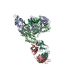
Mass: 24.305 Da / Num. of mol.: 1 / Source method: obtained synthetically / Formula: Mg

-
Experimental details

-
Experiment

ExperimentMethod: X-RAY DIFFRACTION / Number of used crystals: 1

-
Sample preparation

CrystalDensity Matthews: 5.2 Å3/Da / Density % sol: 76.16 %
Crystal growTemperature: 277 K / Method: vapor diffusion, hanging drop / pH: 5.6
Details: cacodylate, SAS, pH 5.6, VAPOR DIFFUSION, HANGING DROP, temperature 277K
Components of the solutions
IDNameCrystal-IDSol-ID
1cacodylateCacodylic acid11
2SAS11
Crystal grow
*PLUS
Temperature: 40 ℃
Components of the solutions
*PLUS
IDConc.Common nameCrystal-IDSol-IDDetails
120 mg/mlprotein1drop
2100 mMcacodylate1reservoirpH5.6
331-34 %satammonium sulfate1reservoir

-
Data collection

DiffractionMean temperature: 105 K
Diffraction sourceSource: SYNCHROTRON / Site: APS / Beamline: 14-BM-C / Wavelength: 1 Å
DetectorType: ADSC QUANTUM 4 / Detector: CCD / Date: Oct 1, 2000
RadiationProtocol: SINGLE WAVELENGTH / Monochromatic (M) / Laue (L): M / Scattering type: x-ray
Radiation wavelengthWavelength: 1 Å / Relative weight: 1
ReflectionResolution: 3.1→35 Å / Num. obs: 61682 / % possible obs: 96.7 % / Observed criterion σ(I): 0 / Redundancy: 5.2 % / Biso Wilson estimate: 61.7 Å2 / Rsym value: 0.11
Reflection shellResolution: 3.1→3.15 Å / Mean I/σ(I) obs: 1.3 / Num. unique all: 2658 / Rsym value: 0.4 / % possible all: 82.9
Reflection
*PLUS
Lowest resolution: 40 Å / % possible obs: 96.7 % / Num. measured all: 930560 / Rmerge(I) obs: 0.11
Reflection shell
*PLUS
% possible obs: 82.7 %

-
Processing

Software
NameVersionClassification
DENZOdata reduction
SCALEPACKdata scaling
X-PLORmodel building
CNS1refinement
X-PLORphasing
RefinementMethod to determine structure: MOLECULAR REPLACEMENT / Resolution: 3.1→20 Å / Rfactor Rfree error: 0.006 / Isotropic thermal model: RESTRAINED / Cross valid method: THROUGHOUT / σ(F): 2 / Stereochemistry target values: Engh & Huber
RfactorNum. reflection% reflectionSelection details
Rfree0.285 2376 4.1 %RANDOM
Rwork0.255 ---
obs0.255 58391 91.8 %-
all-60767 --
Solvent computationSolvent model: FLAT MODEL / Bsol: 27.4085 Å2 / ksol: 0.244335 e/Å3
Displacement parameters
Baniso -1Baniso -2Baniso -3
1-0 Å20 Å20 Å2
2--0 Å20 Å2
3---0 Å2
Refine analyze
FreeObs
Luzzati coordinate error0.5 Å0.45 Å
Refinement stepCycle: LAST / Resolution: 3.1→20 Å
ProteinNucleic acidLigandSolventTotal
Num. atoms11344 840 1 0 12185
Refine LS restraints
Refine-IDTypeDev ideal
X-RAY DIFFRACTIONc_bond_d0.014
X-RAY DIFFRACTIONc_angle_deg1.8
X-RAY DIFFRACTIONc_dihedral_angle_d
X-RAY DIFFRACTIONc_improper_angle_d1.42
LS refinement shellResolution: 3.1→3.21 Å / Rfactor Rfree error: 0.032 / Total num. of bins used: 10
RfactorNum. reflection% reflection
Rfree0.419 172 4 %
Rwork0.399 4100 -
obs--67.6 %
Xplor file
Refine-IDSerial noParam fileTopol file
X-RAY DIFFRACTION1PROTEIN_REP.PARAMPROTEIN.TOP
X-RAY DIFFRACTION2ION.PARAMION.TOP
X-RAY DIFFRACTION3AZT.PARAMAZT.TOP
X-RAY DIFFRACTION4DNA-RNA.PARAMDNA-RNA.TOP
X-RAY DIFFRACTION5&_1_PARAMETER_INFILE_5&_1_TOPOLOGY_INFILE_5
Refinement
*PLUS
Highest resolution: 3.1 Å / Lowest resolution: 20 Å
Solvent computation
*PLUS
Displacement parameters
*PLUS
Refine LS restraints
*PLUS
Refine-IDTypeDev ideal
X-RAY DIFFRACTIONc_improper_angle_d
X-RAY DIFFRACTIONc_improper_angle_deg1.42

+
About Yorodumi

-
News

-
Feb 9, 2022. New format data for meta-information of EMDB entries

New format data for meta-information of EMDB entries

  • Version 3 of the EMDB header file is now the official format.
  • The previous official version 1.9 will be removed from the archive.

Related info.:EMDB header

External links:wwPDB to switch to version 3 of the EMDB data model

-
Aug 12, 2020. Covid-19 info

Covid-19 info

URL: https://pdbj.org/emnavi/covid19.php

New page: Covid-19 featured information page in EM Navigator.

Related info.:Covid-19 info / Mar 5, 2020. Novel coronavirus structure data

+
Mar 5, 2020. Novel coronavirus structure data

Novel coronavirus structure data

Related info.:Yorodumi Speices / Aug 12, 2020. Covid-19 info

External links:COVID-19 featured content - PDBj / Molecule of the Month (242):Coronavirus Proteases

+
Jan 31, 2019. EMDB accession codes are about to change! (news from PDBe EMDB page)

EMDB accession codes are about to change! (news from PDBe EMDB page)

  • The allocation of 4 digits for EMDB accession codes will soon come to an end. Whilst these codes will remain in use, new EMDB accession codes will include an additional digit and will expand incrementally as the available range of codes is exhausted. The current 4-digit format prefixed with “EMD-” (i.e. EMD-XXXX) will advance to a 5-digit format (i.e. EMD-XXXXX), and so on. It is currently estimated that the 4-digit codes will be depleted around Spring 2019, at which point the 5-digit format will come into force.
  • The EM Navigator/Yorodumi systems omit the EMD- prefix.

Related info.:Q: What is EMD? / ID/Accession-code notation in Yorodumi/EM Navigator

External links:EMDB Accession Codes are Changing Soon! / Contact to PDBj

+
Jul 12, 2017. Major update of PDB

Major update of PDB

  • wwPDB released updated PDB data conforming to the new PDBx/mmCIF dictionary.
  • This is a major update changing the version number from 4 to 5, and with Remediation, in which all the entries are updated.
  • In this update, many items about electron microscopy experimental information are reorganized (e.g. em_software).
  • Now, EM Navigator and Yorodumi are based on the updated data.

External links:wwPDB Remediation / Enriched Model Files Conforming to OneDep Data Standards Now Available in the PDB FTP Archive

-
Yorodumi

Thousand views of thousand structures

  • Yorodumi is a browser for structure data from EMDB, PDB, SASBDB, etc.
  • This page is also the successor to EM Navigator detail page, and also detail information page/front-end page for Omokage search.
  • The word "yorodu" (or yorozu) is an old Japanese word meaning "ten thousand". "mi" (miru) is to see.

Related info.:EMDB / PDB / SASBDB / Comparison of 3 databanks / Yorodumi Search / Aug 31, 2016. New EM Navigator & Yorodumi / Yorodumi Papers / Jmol/JSmol / Function and homology information / Changes in new EM Navigator and Yorodumi

Read more