 Movie
Movie Controller

[English] 日本語
 Yorodumi
- PDB-2ban: Crystal structure of HIV-1 reverse transcriptase (RT) in complex ... -
+ Open data

- Basic information
| Entry | Database: PDB / ID: 2ban | ||||||
|---|---|---|---|---|---|---|---|
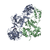
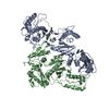
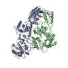
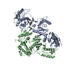
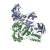
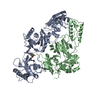
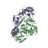
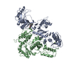
| Title | Crystal structure of HIV-1 reverse transcriptase (RT) in complex with JANSSEN-R157208 | ||||||
 Components Components |
| ||||||
 Keywords Keywords | TRANSFERASE / AIDS / HIV / drug design / reverse transcriptase / RT / protein-inhibitor complex | ||||||
| Function / homology |  Function and homology information Function and homology informationHIV-1 retropepsin / symbiont-mediated activation of host apoptosis / retroviral ribonuclease H / exoribonuclease H / exoribonuclease H activity / DNA integration / viral genome integration into host DNA / establishment of integrated proviral latency / RNA-directed DNA polymerase / RNA stem-loop binding ...HIV-1 retropepsin / symbiont-mediated activation of host apoptosis / retroviral ribonuclease H / exoribonuclease H / exoribonuclease H activity / DNA integration / viral genome integration into host DNA / establishment of integrated proviral latency / RNA-directed DNA polymerase / RNA stem-loop binding / viral penetration into host nucleus / host multivesicular body / RNA-directed DNA polymerase activity / RNA-DNA hybrid ribonuclease activity / Transferases; Transferring phosphorus-containing groups; Nucleotidyltransferases / host cell / viral nucleocapsid / DNA recombination / DNA-directed DNA polymerase / aspartic-type endopeptidase activity / Hydrolases; Acting on ester bonds / DNA-directed DNA polymerase activity / symbiont-mediated suppression of host gene expression / viral translational frameshifting / symbiont entry into host cell / lipid binding / host cell nucleus / host cell plasma membrane / virion membrane / structural molecule activity / proteolysis / DNA binding / zinc ion binding Similarity search - Function | ||||||
| Biological species |   Human immunodeficiency virus 1 Human immunodeficiency virus 1 | ||||||
| Method |  X-RAY DIFFRACTION / SYNCHROTRON / MOLECULAR REPLACEMENT / Resolution: 2.95 Å X-RAY DIFFRACTION / SYNCHROTRON / MOLECULAR REPLACEMENT / Resolution: 2.95 Å | ||||||
 Authors Authors | Das, K. / Arnold, E. | ||||||
 Citation Citation | Journal: J.Med.Chem. / Year: 2005 Title: Crystal Structures for HIV-1 Reverse Transcriptase in Complexes with Three Pyridinone Derivatives: A New Class of Non-Nucleoside Inhibitors Effective against a Broad Range of Drug-Resistant Strains. Authors: Himmel, D.M. / Das, K. / Clark Jr., A.D. / Hughes, S.H. / Benjahad, A. / Oumouch, S. / Guillemont, J. / Coupa, S. / Poncelet, A. / Csoka, I. / Meyer, C. / Andries, K. / Nguyen, C.H. / Grierson, D.S. / Arnold, E. #1: Journal: J.MED.CHEM. / Year: 2005Title: 4-benzyl and 4-benzoyl-3-dimethylaminopyridin-2(1h)-ones: in vitro evaluation of new c-3-amino-substituted and c-5,6-alkyl-substituted analogues against clinically important HIV mutant strains Authors: Benjahad, A. / Croisy, M. / Monneret, C. / Bisagni, E. / Mabire, D. / Coupa, S. / Poncelet, A. / Csoka, I. / Guillemont, J. / Meyer, C. / Andries, K. / Pauwels, R. / de Bethune, M.P. / ...Authors: Benjahad, A. / Croisy, M. / Monneret, C. / Bisagni, E. / Mabire, D. / Coupa, S. / Poncelet, A. / Csoka, I. / Guillemont, J. / Meyer, C. / Andries, K. / Pauwels, R. / de Bethune, M.P. / Himmel, D.M. / Das, K. / Arnold, E. / Nguyen, C.H. / Grierson, D.S. #2: Journal: J.Med.Chem. / Year: 2004Title: Roles of Conformational and Positional Adaptability in Structure-Based Design of TMC125-R165335 (Etravirine) and Related Non-nucleoside Reverse Transcriptase Inhibitors That Are Highly Potent ...Title: Roles of Conformational and Positional Adaptability in Structure-Based Design of TMC125-R165335 (Etravirine) and Related Non-nucleoside Reverse Transcriptase Inhibitors That Are Highly Potent and Effective against Wild-Type and Drug-Resistant HIV-1 Variants Authors: Das, K. / Clark Jr., A.D. / Lewi, P.J. / Heeres, J. / de Jonge, M.R. / Koymans, L.M.H. / Vinkers, H.M. / Daeyaert, F. / Ludovici, D.W. / Kukla, M.J. / De Corte, B. / Kavash, R.W. / Ho, C.Y. ...Authors: Das, K. / Clark Jr., A.D. / Lewi, P.J. / Heeres, J. / de Jonge, M.R. / Koymans, L.M.H. / Vinkers, H.M. / Daeyaert, F. / Ludovici, D.W. / Kukla, M.J. / De Corte, B. / Kavash, R.W. / Ho, C.Y. / Ye, H. / Lichtenstein, M.A. / Andries, K. / Pauwels, R. / de Bethune, M.-P. / Boyer, P.L. / Clark, P. / Hughes, S.H. / Janssen, P.A.J. / Arnold, E. #3: Journal: J.Mol.Biol. / Year: 1996Title: Crystal Structures of 8-Cl and 9-Cl TIBO Complexed with Wild-Type HIV-1 RT and 8-Cl TIBO Complexed with the Tyr181Cys HIV-1 RT Drug-Resistant Mutant. Authors: Das, K. / Ding, J. / Hsiou, Y. / Clark Jr., A.D. / Moereels, H. / Koymans, L. / Andries, K. / Pauwels, R. / Janssen, P.A. / Boyer, P.L. / Clark, P. / Smith Jr., R.H. / Kroeger Smith, M.B. / ...Authors: Das, K. / Ding, J. / Hsiou, Y. / Clark Jr., A.D. / Moereels, H. / Koymans, L. / Andries, K. / Pauwels, R. / Janssen, P.A. / Boyer, P.L. / Clark, P. / Smith Jr., R.H. / Kroeger Smith, M.B. / Michejda, C.J. / Hughes, S.H. / Arnold, E. #4: Journal: J.Mol.Biol. / Year: 1998Title: Structure and Functional Implications of the Polymerase Active Site Region in a Complex of HIV-1 RT with a Double-Stranded DNA Template-Primer and an Antibody Fab Fragment at 2.8 A Resolution. Authors: Ding, J. / Das, K. / Hsiou, Y. / Sarafianos, S.G. / Clark Jr., A.D. / Jacobo-Molina, A. / Tantillo, C. / Hughes, S.H. / Arnold, E. #5: Journal: Nat.Struct.Biol. / Year: 1995Title: Structure of HIV-1 RT/TIBO R 86183 Complex Reveals Similarity in the Binding of Diverse Nonnucleoside Inhibitors. Authors: Ding, J. / Das, K. / Moereels, H. / Koymans, L. / Andries, K. / Janssen, P.A.J. / Hughes, S.H. / Arnold, E. | ||||||
| History |
|
- Structure visualization
| Structure viewer | Molecule: MolmilJmol/JSmol |
|---|
- Downloads & links
-Download
| PDBx/mmCIF format | 2ban.cif.gz | 212.3 KB | Display | PDBx/mmCIF format |
|---|---|---|---|---|
| PDB format | pdb2ban.ent.gz | 166.3 KB | Display | PDB format |
| PDBx/mmJSON format | 2ban.json.gz | Tree view | PDBx/mmJSON format | |
| Others |  Other downloads Other downloads |
-Validation report
| Arichive directory | https://data.pdbj.org/pub/pdb/validation_reports/ba/2banftp://data.pdbj.org/pub/pdb/validation_reports/ba/2ban | HTTPS FTP |
|---|
-Related structure data
| Related structure data |  2b5jC  2be2C  1hnvS C: citing same article ( S: Starting model for refinement |
|---|---|
| Similar structure data |


-Links
 PDBj
PDBj


- Assembly
| Deposited unit | 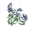
| ||||||||
|---|---|---|---|---|---|---|---|---|---|
| 1 |
| ||||||||
| Unit cell |
|
-Components
| #1: Protein | Mass: 64500.965 Da / Num. of mol.: 1 / Fragment: RESIDUES 599-1158 / Mutation: C447S Source method: isolated from a genetically manipulated source Source: (gene. exp.) Human immunodeficiency virus 1 / Genus: Lentivirus / Strain: BH10 ISOLATE / Description: HIV-1 CLONE 12 / Production host:  |
|---|---|
| #2: Protein | Mass: 50281.762 Da / Num. of mol.: 1 / Fragment: RESIDUES 599-1028 / Mutation: C447S Source method: isolated from a genetically manipulated source Source: (gene. exp.) Human immunodeficiency virus 1 / Genus: Lentivirus / Strain: BH10 ISOLATE / Description: HIV-1 CLONE 12 / Production host: |
| #3: Chemical | ChemComp-MN /   Mass: 54.938 Da / Num. of mol.: 1 / Source method: obtained synthetically / Formula: Mn Mass: 54.938 Da / Num. of mol.: 1 / Source method: obtained synthetically / Formula: Mn |
| #4: Chemical | ChemComp-357 /   Mass: 328.449 Da / Num. of mol.: 1 / Source method: obtained synthetically / Formula: C20H28N2O2 Mass: 328.449 Da / Num. of mol.: 1 / Source method: obtained synthetically / Formula: C20H28N2O2 |
| #5: Water | ChemComp-HOH /  Mass: 18.015 Da / Num. of mol.: 1 / Source method: isolated from a natural source / Formula: H2O Mass: 18.015 Da / Num. of mol.: 1 / Source method: isolated from a natural source / Formula: H2O |
-Experimental details
-Experiment
| Experiment | Method: X-RAY DIFFRACTION / Number of used crystals: 1 |
|---|
- Sample preparation
| Crystal | Density Matthews: 3.42 Å3/Da / Density % sol: 64.08 % |
|---|---|
| Crystal grow | Temperature: 277 K / Method: vapor diffusion, hanging drop / pH: 6.8 Details: PEG8000, AMMINIUM SULPHATE, SODIUM, CHLORID, MANGANESE CHLORIDE, pH 6.8, VAPOR DIFFUSION, HANGING DROP, temperature 277K |
-Data collection
| Diffraction | Mean temperature: 100 K |
|---|---|
| Diffraction source | Source: SYNCHROTRON / Site: APS  / Beamline: 14-BM-D / Wavelength: 1 Å / Beamline: 14-BM-D / Wavelength: 1 Å |
| Detector | Type: ADSC QUANTUM 4 / Detector: CCD / Date: Feb 24, 2001 |
| Radiation | Monochromator: GRAPHITE / Protocol: SINGLE WAVELENGTH / Monochromatic (M) / Laue (L): M / Scattering type: x-ray |
| Radiation wavelength | Wavelength: 1 Å / Relative weight: 1 |
| Reflection | Resolution: 2.95→40 Å / Num. obs: 31625 / % possible obs: 95.5 % / Observed criterion σ(I): -1 / Rmerge(I) obs: 0.063 / Χ2: 1.014 |
| Reflection shell | Resolution: 2.95→3.06 Å / % possible obs: 77.4 % / Rmerge(I) obs: 0.404 / Num. measured obs: 2544 / Χ2: 0.923 / % possible all: 77.4 |
- Processing
| Software |
| ||||||||||||||||||||||||||||||||||||
|---|---|---|---|---|---|---|---|---|---|---|---|---|---|---|---|---|---|---|---|---|---|---|---|---|---|---|---|---|---|---|---|---|---|---|---|---|---|
| Refinement | Method to determine structure: MOLECULAR REPLACEMENT Starting model: PDB entry 1HNV Resolution: 2.95→19.86 Å / Rfactor Rfree error: 0.008 / Data cutoff high absF: 382406.531 / Data cutoff low absF: 0 / Isotropic thermal model: RESTRAINED / Cross valid method: THROUGHOUT / σ(F): 1 / Stereochemistry target values: Engh & Huber
| ||||||||||||||||||||||||||||||||||||
| Solvent computation | Solvent model: FLAT MODEL / Bsol: 14.902 Å2 / ksol: 0.23 e/Å3 | ||||||||||||||||||||||||||||||||||||
| Displacement parameters |
| ||||||||||||||||||||||||||||||||||||
| Refine analyze |
| ||||||||||||||||||||||||||||||||||||
| Refinement step | Cycle: LAST / Resolution: 2.95→19.86 Å
| ||||||||||||||||||||||||||||||||||||
| Refine LS restraints |
| ||||||||||||||||||||||||||||||||||||
| LS refinement shell | Resolution: 2.95→3.13 Å / Rfactor Rfree error: 0.035 / Total num. of bins used: 6
| ||||||||||||||||||||||||||||||||||||
| Xplor file |
|
