 Movie
Movie Controller


+ Open data

- Basic information
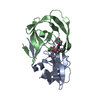
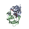
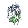
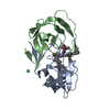
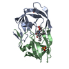
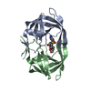
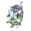
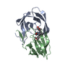
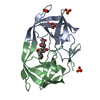
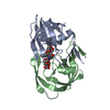
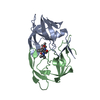
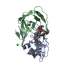
| Entry | Database: PDB / ID: 1meu | ||||||
|---|---|---|---|---|---|---|---|
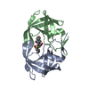
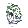
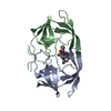
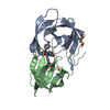
| Title | HIV-1 MUTANT (V82F, I84V) PROTEASE COMPLEXED WITH DMP323 | ||||||
 Components Components | HIV-1 PROTEASE | ||||||
 Keywords Keywords | ASPARTYL PROTEASE / HYDROLASE / ACID PROTEINASE | ||||||
| Function / homology |  Function and homology information Function and homology informationHIV-1 retropepsin / symbiont-mediated activation of host apoptosis / retroviral ribonuclease H / exoribonuclease H / exoribonuclease H activity / DNA integration / viral genome integration into host DNA / establishment of integrated proviral latency / RNA-directed DNA polymerase / RNA stem-loop binding ...HIV-1 retropepsin / symbiont-mediated activation of host apoptosis / retroviral ribonuclease H / exoribonuclease H / exoribonuclease H activity / DNA integration / viral genome integration into host DNA / establishment of integrated proviral latency / RNA-directed DNA polymerase / RNA stem-loop binding / viral penetration into host nucleus / host multivesicular body / RNA-directed DNA polymerase activity / RNA-DNA hybrid ribonuclease activity / Transferases; Transferring phosphorus-containing groups; Nucleotidyltransferases / host cell / viral nucleocapsid / DNA recombination / DNA-directed DNA polymerase / aspartic-type endopeptidase activity / Hydrolases; Acting on ester bonds / DNA-directed DNA polymerase activity / symbiont-mediated suppression of host gene expression / viral translational frameshifting / symbiont entry into host cell / lipid binding / host cell nucleus / host cell plasma membrane / virion membrane / structural molecule activity / proteolysis / DNA binding / zinc ion binding Similarity search - Function | ||||||
| Biological species |   Human immunodeficiency virus 1 Human immunodeficiency virus 1 | ||||||
| Method |  X-RAY DIFFRACTION / Resolution: 1.9 Å X-RAY DIFFRACTION / Resolution: 1.9 Å | ||||||
 Authors Authors | Ala, P. / Chang, C.-H. | ||||||
 Citation Citation | Journal: Biochemistry / Year: 1997 Title: Molecular basis of HIV-1 protease drug resistance: structural analysis of mutant proteases complexed with cyclic urea inhibitors. Authors: Ala, P.J. / Huston, E.E. / Klabe, R.M. / McCabe, D.D. / Duke, J.L. / Rizzo, C.J. / Korant, B.D. / DeLoskey, R.J. / Lam, P.Y. / Hodge, C.N. / Chang, C.H. #1: Journal: Biochemistry / Year: 1997Title: Erratum. Molecular Basis of HIV-1 Protease Drug Resistance: Structural Analysis of Mutant Proteases Complexed with Cyclic Urea Inhibitors Authors: Ala, P.J. / Huston, E.E. / Klabe, R.M. / Mccabe, D.D. / Duke, J.L. / Rizzo, C.J. / Korant, B.D. / Deloskey, R.J. / Lam, P.Y. / Hodge, C.N. / Chang, C.H. | ||||||
| History |
|
- Structure visualization
| Structure viewer | Molecule: MolmilJmol/JSmol |
|---|
- Downloads & links
-Download
| PDBx/mmCIF format | 1meu.cif.gz | 57 KB | Display | PDBx/mmCIF format |
|---|---|---|---|---|
| PDB format | pdb1meu.ent.gz | 41.9 KB | Display | PDB format |
| PDBx/mmJSON format | 1meu.json.gz | Tree view | PDBx/mmJSON format | |
| Others |  Other downloads Other downloads |
-Validation report
| Arichive directory | https://data.pdbj.org/pub/pdb/validation_reports/me/1meuftp://data.pdbj.org/pub/pdb/validation_reports/me/1meu | HTTPS FTP |
|---|
-Related structure data
| Related structure data | 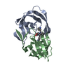 1merC 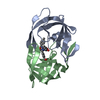 1mesC 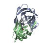 1metC 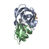 1hvrS S: Starting model for refinement C: citing same article ( |
|---|---|
| Similar structure data |


-Links
 PDBj
PDBj


- Assembly
| Deposited unit | 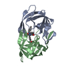
| ||||||||
|---|---|---|---|---|---|---|---|---|---|
| 1 |
| ||||||||
| Unit cell |
|
-Components
| #1: Protein | Mass: 10823.746 Da / Num. of mol.: 2 / Mutation: V82F, I84V Source method: isolated from a genetically manipulated source Source: (gene. exp.) Human immunodeficiency virus 1 / Genus: Lentivirus / Strain: BH102 ISOLATE / Production host:  #2: Chemical | ChemComp-DMP / [ |   Mass: 566.687 Da / Num. of mol.: 1 / Source method: obtained synthetically / Formula: C35H38N2O5 Mass: 566.687 Da / Num. of mol.: 1 / Source method: obtained synthetically / Formula: C35H38N2O5 |
|---|
-Experimental details
-Experiment
| Experiment | Method: X-RAY DIFFRACTION |
|---|
- Sample preparation
| Crystal | Density Matthews: 2.19 Å3/Da / Density % sol: 43.95 % | ||||||||||||||||||||
|---|---|---|---|---|---|---|---|---|---|---|---|---|---|---|---|---|---|---|---|---|---|
| Crystal grow | *PLUS Temperature: 18 ℃ / Method: vapor diffusion, hanging drop / PH range low: 5.6 / PH range high: 5 | ||||||||||||||||||||
| Components of the solutions | *PLUS
|
-Data collection
| Radiation | Scattering type: x-ray |
|---|---|
| Radiation wavelength | Relative weight: 1 |
| Reflection | *PLUS Highest resolution: 1.9 Å / Num. obs: 14386 / % possible obs: 89.6 % / Num. measured all: 41114 / Rmerge(I) obs: 0.088 |
- Processing
| Software |
| ||||||||||||||||||||||||||||||||||||||||||||||||||||||||||||
|---|---|---|---|---|---|---|---|---|---|---|---|---|---|---|---|---|---|---|---|---|---|---|---|---|---|---|---|---|---|---|---|---|---|---|---|---|---|---|---|---|---|---|---|---|---|---|---|---|---|---|---|---|---|---|---|---|---|---|---|---|---|
| Refinement | Starting model: PDB ENTRY 1HVR Rfactor Rfree: 0.193 / Highest resolution: 1.9 Å | ||||||||||||||||||||||||||||||||||||||||||||||||||||||||||||
| Refinement step | Cycle: LAST / Highest resolution: 1.9 Å
| ||||||||||||||||||||||||||||||||||||||||||||||||||||||||||||
| Refine LS restraints |
| ||||||||||||||||||||||||||||||||||||||||||||||||||||||||||||
| Refinement | *PLUS σ(F): 2 / Rfactor obs: 0.193 | ||||||||||||||||||||||||||||||||||||||||||||||||||||||||||||
| Solvent computation | *PLUS | ||||||||||||||||||||||||||||||||||||||||||||||||||||||||||||
| Displacement parameters | *PLUS |
