 Movie
Movie Controller

[English] 日本語
 Yorodumi
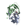
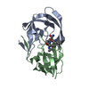
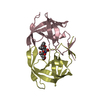
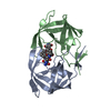
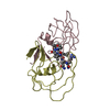
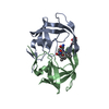
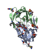
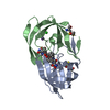
- PDB-1bdq: HIV-1 (2:31-37, 47, 82) PROTEASE COMPLEXED WITH INHIBITOR SB203386 -
+ Open data

- Basic information
| Entry | Database: PDB / ID: 1bdq | ||||||
|---|---|---|---|---|---|---|---|
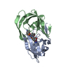
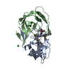
| Title | HIV-1 (2:31-37, 47, 82) PROTEASE COMPLEXED WITH INHIBITOR SB203386 | ||||||
 Components Components | HIV-1 PROTEASE | ||||||
 Keywords Keywords | HYDROLASE / AIDS / POLYPROTEIN / ASPARTYL PROTEASE / ACID PROTEASE / HYDROXYETHYLENE ISOSTERE INHIBITOR / SUBSTRATE ANALOGUE INHIBITOR | ||||||
| Function / homology |  Function and homology information Function and homology informationHIV-1 retropepsin / symbiont-mediated activation of host apoptosis / retroviral ribonuclease H / exoribonuclease H / exoribonuclease H activity / DNA integration / viral genome integration into host DNA / establishment of integrated proviral latency / RNA-directed DNA polymerase / RNA stem-loop binding ...HIV-1 retropepsin / symbiont-mediated activation of host apoptosis / retroviral ribonuclease H / exoribonuclease H / exoribonuclease H activity / DNA integration / viral genome integration into host DNA / establishment of integrated proviral latency / RNA-directed DNA polymerase / RNA stem-loop binding / viral penetration into host nucleus / host multivesicular body / RNA-directed DNA polymerase activity / RNA-DNA hybrid ribonuclease activity / Transferases; Transferring phosphorus-containing groups; Nucleotidyltransferases / host cell / viral nucleocapsid / DNA recombination / DNA-directed DNA polymerase / aspartic-type endopeptidase activity / Hydrolases; Acting on ester bonds / DNA-directed DNA polymerase activity / symbiont-mediated suppression of host gene expression / viral translational frameshifting / symbiont entry into host cell / lipid binding / host cell nucleus / host cell plasma membrane / virion membrane / structural molecule activity / proteolysis / DNA binding / zinc ion binding Similarity search - Function | ||||||
| Biological species |   Human immunodeficiency virus 1 Human immunodeficiency virus 1 | ||||||
| Method |  X-RAY DIFFRACTION / DIFFERENCE FOURIER / Resolution: 2.5 Å X-RAY DIFFRACTION / DIFFERENCE FOURIER / Resolution: 2.5 Å | ||||||
 Authors Authors | Swairjo, M.A. / Abdel-Meguid, S.S. | ||||||
 Citation Citation | Journal: Biochemistry / Year: 1998 Title: Structural role of the 30's loop in determining the ligand specificity of the human immunodeficiency virus protease. Authors: Swairjo, M.A. / Towler, E.M. / Debouck, C. / Abdel-Meguid, S.S. #1: Journal: Biochemistry / Year: 1997Title: Identification of a Loop Outside the Active Site Cavity of the Human Immunodeficiency Virus Proteases which Confers Inhibitor Specificity Authors: Towler, E.M. / Thompson, S.K. / Tomaszek, T. / Debouck, C. | ||||||
| History |
|
- Structure visualization
| Structure viewer | Molecule: MolmilJmol/JSmol |
|---|
- Downloads & links
-Download
| PDBx/mmCIF format | 1bdq.cif.gz | 48.8 KB | Display | PDBx/mmCIF format |
|---|---|---|---|---|
| PDB format | pdb1bdq.ent.gz | 34.5 KB | Display | PDB format |
| PDBx/mmJSON format | 1bdq.json.gz | Tree view | PDBx/mmJSON format | |
| Others |  Other downloads Other downloads |
-Validation report
| Arichive directory | https://data.pdbj.org/pub/pdb/validation_reports/bd/1bdqftp://data.pdbj.org/pub/pdb/validation_reports/bd/1bdq | HTTPS FTP |
|---|
-Related structure data
| Related structure data |  1bdlC  1bdrC  1sbgS S: Starting model for refinement C: citing same article ( |
|---|---|
| Similar structure data |

-Links
 PDBj
PDBj
- Assembly
| Deposited unit | 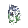
| ||||||||
|---|---|---|---|---|---|---|---|---|---|
| 1 |
| ||||||||
| Unit cell |
|
-Components
| #1: Protein | Mass: 10683.630 Da / Num. of mol.: 2 Mutation: T31S, V32I, L33V, E34A, E35G, M36I, S37E, I47V, V82I Source method: isolated from a genetically manipulated source Source: (gene. exp.) Human immunodeficiency virus 1 / Genus: Lentivirus / Strain: IIIB / Gene: HIV-1 PROTEASE / Plasmid: POTSKF33 / Gene (production host): HIV-1 PROTEASE / Production host:  #2: Chemical | ChemComp-IM1 / ( |   Mass: 534.690 Da / Num. of mol.: 1 / Source method: obtained synthetically / Formula: C31H42N4O4 Mass: 534.690 Da / Num. of mol.: 1 / Source method: obtained synthetically / Formula: C31H42N4O4#3: Water | ChemComp-HOH / |  Mass: 18.015 Da / Num. of mol.: 43 / Source method: isolated from a natural source / Formula: H2O Mass: 18.015 Da / Num. of mol.: 43 / Source method: isolated from a natural source / Formula: H2O |
|---|
-Experimental details
-Experiment
| Experiment | Method: X-RAY DIFFRACTION / Number of used crystals: 1 |
|---|
- Sample preparation
| Crystal | Density Matthews: 2.22 Å3/Da / Density % sol: 45 % | ||||||||||||||||||||||||||||||||||||||||||||||||
|---|---|---|---|---|---|---|---|---|---|---|---|---|---|---|---|---|---|---|---|---|---|---|---|---|---|---|---|---|---|---|---|---|---|---|---|---|---|---|---|---|---|---|---|---|---|---|---|---|---|
| Crystal grow | Temperature: 294 K / Method: vapor diffusion, hanging drop / pH: 5.6 Details: HANGING DROP VAPOUR DIFFUSION, BY MIXING EQUAL VOLUMES OF RESERVOIR AND SAMPLE, 21 DEGREES C. RESERVOIR: 35% 3.5 MG/ML PROTEIN/INHIBITOR COMPLEX AT 1:5 MOLAR RATIO., pH 5.6, vapor diffusion - ...Details: HANGING DROP VAPOUR DIFFUSION, BY MIXING EQUAL VOLUMES OF RESERVOIR AND SAMPLE, 21 DEGREES C. RESERVOIR: 35% 3.5 MG/ML PROTEIN/INHIBITOR COMPLEX AT 1:5 MOLAR RATIO., pH 5.6, vapor diffusion - hanging drop, temperature 294K | ||||||||||||||||||||||||||||||||||||||||||||||||
| Crystal | *PLUS | ||||||||||||||||||||||||||||||||||||||||||||||||
| Crystal grow | *PLUS Temperature: 21 ℃ / pH: 5 / Method: vapor diffusion, hanging drop | ||||||||||||||||||||||||||||||||||||||||||||||||
| Components of the solutions | *PLUS
|
-Data collection
| Diffraction | Mean temperature: 292 K |
|---|---|
| Diffraction source | Source: ROTATING ANODE / Type: RIGAKU RUH2R / Wavelength: 1.5418 |
| Detector | Type: MARRESEARCH / Detector: IMAGE PLATE / Date: Nov 1, 1997 / Details: MIRRORS |
| Radiation | Monochromator: NI FILTER / Monochromatic (M) / Laue (L): M / Scattering type: x-ray |
| Radiation wavelength | Wavelength: 1.5418 Å / Relative weight: 1 |
| Reflection | Resolution: 2.5→20 Å / Num. obs: 5858 / % possible obs: 99 % / Observed criterion σ(I): 1 / Redundancy: 3.1 % / Rmerge(I) obs: 0.14 / Rsym value: 0.14 / Net I/σ(I): 14.1 |
| Reflection shell | Resolution: 2.5→2.59 Å / Redundancy: 3 % / Rmerge(I) obs: 0.7 / Mean I/σ(I) obs: 6.4 / Rsym value: 0.08 / % possible all: 53.7 |
| Reflection | *PLUS Num. measured all: 43218 |
| Reflection shell | *PLUS % possible obs: 53.7 % |
- Processing
| Software |
| ||||||||||||||||||||||||||||||||||||||||||||||||||||||||||||
|---|---|---|---|---|---|---|---|---|---|---|---|---|---|---|---|---|---|---|---|---|---|---|---|---|---|---|---|---|---|---|---|---|---|---|---|---|---|---|---|---|---|---|---|---|---|---|---|---|---|---|---|---|---|---|---|---|---|---|---|---|---|
| Refinement | Method to determine structure: DIFFERENCE FOURIER Starting model: PDB ENTRY 1SBG Resolution: 2.5→20 Å / Rfactor Rfree error: 0.02 / Data cutoff low absF: 0 / Isotropic thermal model: RESTRAINED / Cross valid method: THROUGHOUT / σ(F): 0
| ||||||||||||||||||||||||||||||||||||||||||||||||||||||||||||
| Displacement parameters | Biso mean: 19.1 Å2
| ||||||||||||||||||||||||||||||||||||||||||||||||||||||||||||
| Refine analyze | Luzzati coordinate error obs: 0.25 Å | ||||||||||||||||||||||||||||||||||||||||||||||||||||||||||||
| Refinement step | Cycle: LAST / Resolution: 2.5→20 Å
| ||||||||||||||||||||||||||||||||||||||||||||||||||||||||||||
| Refine LS restraints |
| ||||||||||||||||||||||||||||||||||||||||||||||||||||||||||||
| LS refinement shell | Resolution: 2.5→2.56 Å / Total num. of bins used: 8 /
| ||||||||||||||||||||||||||||||||||||||||||||||||||||||||||||
| Xplor file |
| ||||||||||||||||||||||||||||||||||||||||||||||||||||||||||||
| Software | *PLUS Name: X-PLOR / Version: 3.1 / Classification: refinement | ||||||||||||||||||||||||||||||||||||||||||||||||||||||||||||
| Refine LS restraints | *PLUS
|
