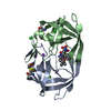
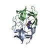
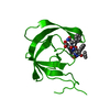
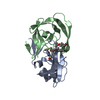
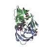
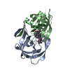
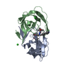
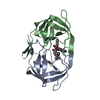
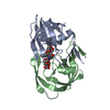
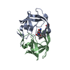
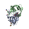
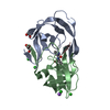
登録情報 データベース : PDB / ID : 1zpkタイトル Crystal structure of the complex of mutant HIV-1 protease (A71V, V82T, I84V) with a hydroxyethylamine peptidomimetic inhibitor BOC-PHE-PSI[R-CH(OH)CH2NH]-PHE-GLU-PHE-NH2 PROTEASE RETROPEPSIN キーワード / / / / 機能・相同性 分子機能 ドメイン・相同性 構成要素
/ / / / / / / / / / / / / / / / / / / / / / / / / / / / / / / / / / / / / / / / / / / / / / / / / / / / / / / / / / / / / / / / / / / / / / / / / / / / / / / / / / / / / / / / / / / / / / / / / / / / / / 生物種 手法 / / / 解像度 : 1.65 Å データ登録者 Duskova, J. / Skalova, T. / Dohnalek, J. / Petrokova, H. / Hasek, J. ジャーナル : To be Published タイトル : Mutational Study of Pseudopeptide Inhibitor Binding to HIV-1 Protease; Analysis of Four X-ray Structures著者 : Duskova, J. / Skalova, T. / Dohnalek, J. / Petrokova, H. / Hasek, J. 履歴 登録 2005年5月17日 登録サイト / 処理サイト 改定 1.0 2006年4月25日 Provider / タイプ 改定 1.1 2007年10月16日 Group 改定 1.2 2011年7月13日 Group Atomic model / Database references ... Atomic model / Database references / Derived calculations / Non-polymer description / Structure summary / Version format compliance 改定 1.3 2012年12月12日 Group 改定 1.4 2021年10月20日 Group / Derived calculationsカテゴリ database_2 / pdbx_struct_conn_angle ... database_2 / pdbx_struct_conn_angle / struct_conn / struct_conn_type / struct_ref_seq_dif / struct_sheet / struct_site Item _database_2.pdbx_DOI / _database_2.pdbx_database_accession ... _database_2.pdbx_DOI / _database_2.pdbx_database_accession / _pdbx_struct_conn_angle.ptnr1_auth_seq_id / _pdbx_struct_conn_angle.ptnr3_auth_seq_id / _pdbx_struct_conn_angle.value / _struct_conn.conn_type_id / _struct_conn.id / _struct_conn.pdbx_dist_value / _struct_conn.pdbx_leaving_atom_flag / _struct_conn.ptnr1_auth_asym_id / _struct_conn.ptnr1_auth_comp_id / _struct_conn.ptnr1_auth_seq_id / _struct_conn.ptnr1_label_asym_id / _struct_conn.ptnr1_label_atom_id / _struct_conn.ptnr1_label_comp_id / _struct_conn.ptnr1_label_seq_id / _struct_conn.ptnr2_auth_asym_id / _struct_conn.ptnr2_auth_comp_id / _struct_conn.ptnr2_auth_seq_id / _struct_conn.ptnr2_label_asym_id / _struct_conn.ptnr2_label_atom_id / _struct_conn.ptnr2_label_comp_id / _struct_conn.ptnr2_label_seq_id / _struct_conn_type.id / _struct_ref_seq_dif.details / _struct_sheet.number_strands / _struct_site.pdbx_auth_asym_id / _struct_site.pdbx_auth_comp_id / _struct_site.pdbx_auth_seq_id 改定 1.5 2023年8月23日 Group / Refinement descriptionカテゴリ / chem_comp_bond / pdbx_initial_refinement_model改定 1.6 2024年11月6日 Group カテゴリ / pdbx_modification_featureItem
すべて表示 表示を減らす
 ムービー
ムービー コントローラー


 データを開く
 基本情報
基本情報 要素
要素 キーワード
キーワード 機能・相同性情報
 Human immunodeficiency virus 1 (ヒト免疫不全ウイルス)
 X線回折 /
  データ登録者
 引用
引用 構造の表示
構造の表示 ダウンロードとリンク
 その他のダウンロード
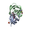


 PDBj
PDBj


 集合体
集合体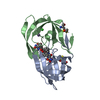

 タイプ: peptide-like, Peptide-like / クラス: 阻害剤 / 分子量: 703.824 Da / 分子数: 1 / 由来タイプ: 合成 / 式: C38H49N5O8 / 参照: BOC-PHE-PSI[R-CH(OH)CH2NH]-PHE-GLU-PHE-NH2
 分子量: 35.453 Da / 分子数: 1 / 由来タイプ: 合成 / 式: Cl
 分子量: 22.990 Da / 分子数: 1 / 由来タイプ: 合成 / 式: Na
 分子量: 18.015 Da / 分子数: 160 / 由来タイプ: 天然 / 式: H2O
 試料調製
試料調製 / ビームライン: ID14-1 / 波長: 0.934 Å
 解析
解析