[English] 日本語
Yorodumi
- PDB-1zj7: Crystal structure of a complex of mutant HIV-1 protease (A71V, V8... -

+
Open data


ID or keywords:

Loading...

-
Basic information

Entry
Database: PDB / ID: 1zj7
Title
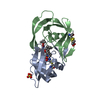
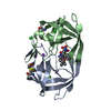
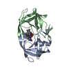
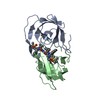
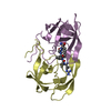
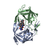
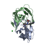
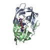
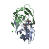
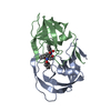
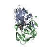
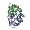
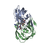
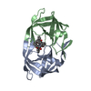
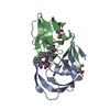
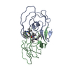
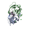
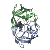
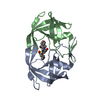
Crystal structure of a complex of mutant HIV-1 protease (A71V, V82T, I84V) with a hydroxyethylamine peptidomimetic inhibitor BOC-PHE-PSI[S-CH(OH)CH2NH]-PHE-GLU-PHE-NH2
ComponentsPROTEASE RETROPEPSIN
KeywordsHYDROLASE/HYDROLASE INHIBITOR / HIV / PROTEASE / PEPTIDOMIMETIC INHIBITOR / HYDROLASE-HYDROLASE INHIBITOR COMPLEX
Function / homology
Function and homology information


RNA stem-loop binding / host cell / HIV-1 retropepsin / retroviral ribonuclease H / exoribonuclease H / exoribonuclease H activity / host multivesicular body / DNA integration / viral genome integration into host DNA / RNA-directed DNA polymerase ...RNA stem-loop binding / host cell / HIV-1 retropepsin / retroviral ribonuclease H / exoribonuclease H / exoribonuclease H activity / host multivesicular body / DNA integration / viral genome integration into host DNA / RNA-directed DNA polymerase / viral penetration into host nucleus / establishment of integrated proviral latency / RNA-directed DNA polymerase activity / RNA-DNA hybrid ribonuclease activity / Transferases; Transferring phosphorus-containing groups; Nucleotidyltransferases / symbiont-mediated suppression of host gene expression / viral nucleocapsid / DNA recombination / DNA-directed DNA polymerase / Hydrolases; Acting on ester bonds / aspartic-type endopeptidase activity / DNA-directed DNA polymerase activity / symbiont entry into host cell / lipid binding / host cell nucleus / host cell plasma membrane / virion membrane / structural molecule activity / proteolysis / DNA binding / zinc ion binding / membrane
Similarity search - Function
Reverse transcriptase connection / Reverse transcriptase connection domain / Reverse transcriptase thumb / Reverse transcriptase thumb domain / Integrase Zinc binding domain / Zinc finger integrase-type profile. / Integrase-like, N-terminal / Integrase DNA binding domain / Integrase, C-terminal domain superfamily, retroviral / Integrase, N-terminal zinc-binding domain ...Reverse transcriptase connection / Reverse transcriptase connection domain / Reverse transcriptase thumb / Reverse transcriptase thumb domain / Integrase Zinc binding domain / Zinc finger integrase-type profile. / Integrase-like, N-terminal / Integrase DNA binding domain / Integrase, C-terminal domain superfamily, retroviral / Integrase, N-terminal zinc-binding domain / Integrase, C-terminal, retroviral / Integrase DNA binding domain profile. / Immunodeficiency lentiviral matrix, N-terminal / gag gene protein p17 (matrix protein) / RNase H / Integrase core domain / Integrase, catalytic core / Integrase catalytic domain profile. / Retroviral nucleocapsid Gag protein p24, C-terminal domain / Gag protein p24 C-terminal domain / Retropepsin-like catalytic domain / Matrix protein, lentiviral and alpha-retroviral, N-terminal / Ribonuclease H domain / RNase H type-1 domain profile. / Reverse transcriptase (RNA-dependent DNA polymerase) / Reverse transcriptase domain / Reverse transcriptase (RT) catalytic domain profile. / Retropepsins / Retroviral aspartyl protease / Aspartyl protease, retroviral-type family profile. / Peptidase A2A, retrovirus, catalytic / Retrovirus capsid, C-terminal / Retroviral matrix protein / Retrovirus capsid, N-terminal / zinc finger / Zinc knuckle / Zinc finger, CCHC-type superfamily / Cathepsin D, subunit A; domain 1 / Acid Proteases / Zinc finger, CCHC-type / Zinc finger CCHC-type profile. / Aspartic peptidase, active site / Eukaryotic and viral aspartyl proteases active site. / Ribonuclease H superfamily / Aspartic peptidase domain superfamily / Ribonuclease H-like superfamily / Reverse transcriptase/Diguanylate cyclase domain / DNA/RNA polymerase superfamily / Beta Barrel / Mainly Beta
Similarity search - Domain/homology
BOC-PHE-PSI[S-CH(OH)CH2NH]-PHE-GLU-PHE-NH2 / Chem-0ZT / Gag-Pol polyprotein
Similarity search - Component
Biological speciesHuman immunodeficiency virus 1
MethodX-RAY DIFFRACTION / SYNCHROTRON / MOLECULAR REPLACEMENT / Resolution: 1.93 Å
AuthorsSkalova, T. / Dohnalek, J. / Duskova, J. / Petrokova, H. / Hasek, J.
CitationJournal: J.Med.Chem. / Year: 2006
Title: HIV-1 protease mutations and inhibitor modifications monitored on a series of complexes. Structural basis for the effect of the A71V mutation on the active site
Authors: Skalova, T. / Dohnalek, J. / Duskova, J. / Petrokova, H. / Hradilek, M. / Soucek, M. / Konvalinka, J. / Hasek, J.
History
DepositionApr 28, 2005Deposition site: RCSB / Processing site: RCSB
Revision 1.0May 9, 2006Provider: repository / Type: Initial release
Revision 1.1Oct 16, 2007Group: Version format compliance
Revision 1.2Jul 13, 2011Group: Atomic model / Database references ...Atomic model / Database references / Derived calculations / Non-polymer description / Structure summary / Version format compliance
Revision 1.3Nov 16, 2011Group: Atomic model
Revision 1.4Dec 12, 2012Group: Other
Revision 1.5Oct 20, 2021Group: Database references / Derived calculations
Category: database_2 / struct_ref_seq_dif ...database_2 / struct_ref_seq_dif / struct_sheet / struct_site
Item: _database_2.pdbx_DOI / _database_2.pdbx_database_accession ..._database_2.pdbx_DOI / _database_2.pdbx_database_accession / _struct_ref_seq_dif.details / _struct_sheet.number_strands / _struct_site.pdbx_auth_asym_id / _struct_site.pdbx_auth_comp_id / _struct_site.pdbx_auth_seq_id
Revision 1.6Aug 23, 2023Group: Data collection / Refinement description
Category: chem_comp_atom / chem_comp_bond / pdbx_initial_refinement_model

-
Structure visualization

Structure viewerMolecule:
MolmilJmol/JSmol

Downloads & links

-
Assembly

Deposited unit
A: PROTEASE RETROPEPSIN
hetero molecules


Theoretical massNumber of molelcules
Total (without water)11,5242
Polymers10,8201
Non-polymers7041
Water1,11762
1
A: PROTEASE RETROPEPSIN
hetero molecules

A: PROTEASE RETROPEPSIN
hetero molecules


Theoretical massNumber of molelcules
Total (without water)23,0474
Polymers21,6402
Non-polymers1,4082
Water362
TypeNameSymmetry operationNumber
identity operation1_555x,y,z1
crystal symmetry operation9_555-x,-x+y,-z+2/31
Buried area7180 Å2
ΔGint-29 kcal/mol
Surface area8860 Å2
MethodPISA
Unit cell
Length a, b, c (Å)62.859, 62.859, 83.253
Angle α, β, γ (deg.)90.00, 90.00, 120.00
Int Tables number178
Cell settinghexagonal
Space group name H-MP6122
Components on special symmetry positions
IDModelComponents
11A-301-

HOH

-
Components

#1: Protein PROTEASE RETROPEPSIN / E.C.3.4.23.16 / HIV-1 PROTEASE


Mass: 10819.756 Da / Num. of mol.: 1 / Mutation: A71V, V82T, I84V
Source method: isolated from a genetically manipulated source
Source: (gene. exp.) Human immunodeficiency virus 1 / Genus: Lentivirus / Plasmid: pET24a / Species (production host): Escherichia coli / Production host: Escherichia coli BL21(DE3) (bacteria) / Strain (production host): BL21(DE3) / References: UniProt: P03367, HIV-1 retropepsin
#2: Chemical ChemComp-0ZT / N-{(2S,3S)-3-[(tert-butoxycarbonyl)amino]-2-hydroxy-4-phenylbutyl}-L-phenylalanyl-L-alpha-glutamyl-L-phenylalaninamide


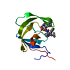
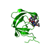
Type: peptide-like, Peptide-like / Class: Inhibitor / Mass: 703.824 Da / Num. of mol.: 1 / Source method: obtained synthetically / Formula: C38H49N5O8 / References: BOC-PHE-PSI[S-CH(OH)CH2NH]-PHE-GLU-PHE-NH2
#3: Water ChemComp-HOH / water


Mass: 18.015 Da / Num. of mol.: 62 / Source method: isolated from a natural source / Formula: H2O

-
Experimental details

-
Experiment

ExperimentMethod: X-RAY DIFFRACTION / Number of used crystals: 1

-
Sample preparation

CrystalDensity Matthews: 1.94 Å3/Da / Density % sol: 36.11 %
Crystal growTemperature: 298 K / Method: vapor diffusion, hanging drop / pH: 4.4
Details: 0.1 M Na Citrate, 0.5 M NaCl, 10% (v/v) glycerol, pH 4.4, VAPOR DIFFUSION, HANGING DROP, temperature 298K

-
Data collection

DiffractionMean temperature: 100 K
Diffraction sourceSource: SYNCHROTRON / Site: ESRF / Beamline: ID14-1 / Wavelength: 0.934 Å
DetectorType: MARRESEARCH / Detector: CCD / Date: Apr 12, 2000 / Details: microfocusing mirrors - testing
RadiationMonochromator: Diamond C 111 / Protocol: SINGLE WAVELENGTH / Monochromatic (M) / Laue (L): M / Scattering type: x-ray
Radiation wavelengthWavelength: 0.934 Å / Relative weight: 1
ReflectionResolution: 1.93→20 Å / Num. all: 6784 / Num. obs: 6784 / % possible obs: 87.6 % / Observed criterion σ(I): -999 / Redundancy: 6.7 % / Biso Wilson estimate: 36.7 Å2 / Rmerge(I) obs: 0.048 / Rsym value: 0.048 / Net I/σ(I): 28.1
Reflection shellResolution: 1.93→2 Å / Redundancy: 6.5 % / Rmerge(I) obs: 0.434 / Mean I/σ(I) obs: 4.4 / Num. unique all: 627 / Rsym value: 0.434 / % possible all: 85.4

-
Processing

Software
NameClassification
DENZOdata reduction
SCALEPACKdata scaling
AMoREphasing
CNSrefinement
RefinementMethod to determine structure: MOLECULAR REPLACEMENT
Starting model: 1IIQ
Resolution: 1.93→20 Å / Data cutoff high absF: 10000 / Data cutoff low absF: 0 / Isotropic thermal model: Isotropic / Cross valid method: THROUGHOUT / σ(F): 0 / Stereochemistry target values: Engh & Huber
Details: Used maximum likelihood refinement procedure on amplitudes and least squares residual on amplitudes.
RfactorNum. reflection% reflectionSelection details
Rfree0.2994 355 5.3 %RANDOM
Rwork0.2288 ---
all0.2287 6714 --
obs0.2287 6714 86.41 %-
Solvent computationSolvent model: AS IMPLEMENTED IN CNS 1.1 / Bsol: 78.09 Å2 / ksol: 0.337 e/Å3
Displacement parametersBiso mean: 33.7 Å2
Baniso -1Baniso -2Baniso -3
1-0.046 Å22.091 Å20 Å2
2--0.046 Å20 Å2
3----0.092 Å2
Refine analyzeLuzzati coordinate error obs: 0.307 Å / Luzzati d res low obs: 5 Å
Refinement stepCycle: LAST / Resolution: 1.93→20 Å
ProteinNucleic acidLigandSolventTotal
Num. atoms759 0 51 62 872
Refine LS restraints
Refine-IDTypeDev idealDev ideal target
X-RAY DIFFRACTIONc_bond_d0.012411
X-RAY DIFFRACTIONc_angle_deg1.70665
X-RAY DIFFRACTIONc_mcbond_it2.3162
X-RAY DIFFRACTIONc_scbond_it3.4423
X-RAY DIFFRACTIONc_mcangle_it3.8814
X-RAY DIFFRACTIONc_scangle_it5.2875
LS refinement shellResolution: 1.93→2.02 Å
RfactorNum. reflection
Rfree0.371 32
Rwork0.3574 -
obs-743

+
About Yorodumi

-
News

-
Feb 9, 2022. New format data for meta-information of EMDB entries

New format data for meta-information of EMDB entries

  • Version 3 of the EMDB header file is now the official format.
  • The previous official version 1.9 will be removed from the archive.

Related info.:EMDB header

External links:wwPDB to switch to version 3 of the EMDB data model

-
Aug 12, 2020. Covid-19 info

Covid-19 info

URL: https://pdbjlvh1.pdbj.org/emnavi/covid19.php

New page: Covid-19 featured information page in EM Navigator.

Related info.:Covid-19 info / Mar 5, 2020. Novel coronavirus structure data

+
Mar 5, 2020. Novel coronavirus structure data

Novel coronavirus structure data

Related info.:Yorodumi Speices / Aug 12, 2020. Covid-19 info

External links:COVID-19 featured content - PDBj / Molecule of the Month (242):Coronavirus Proteases

+
Jan 31, 2019. EMDB accession codes are about to change! (news from PDBe EMDB page)

EMDB accession codes are about to change! (news from PDBe EMDB page)

  • The allocation of 4 digits for EMDB accession codes will soon come to an end. Whilst these codes will remain in use, new EMDB accession codes will include an additional digit and will expand incrementally as the available range of codes is exhausted. The current 4-digit format prefixed with “EMD-” (i.e. EMD-XXXX) will advance to a 5-digit format (i.e. EMD-XXXXX), and so on. It is currently estimated that the 4-digit codes will be depleted around Spring 2019, at which point the 5-digit format will come into force.
  • The EM Navigator/Yorodumi systems omit the EMD- prefix.

Related info.:Q: What is EMD? / ID/Accession-code notation in Yorodumi/EM Navigator

External links:EMDB Accession Codes are Changing Soon! / Contact to PDBj

+
Jul 12, 2017. Major update of PDB

Major update of PDB

  • wwPDB released updated PDB data conforming to the new PDBx/mmCIF dictionary.
  • This is a major update changing the version number from 4 to 5, and with Remediation, in which all the entries are updated.
  • In this update, many items about electron microscopy experimental information are reorganized (e.g. em_software).
  • Now, EM Navigator and Yorodumi are based on the updated data.

External links:wwPDB Remediation / Enriched Model Files Conforming to OneDep Data Standards Now Available in the PDB FTP Archive

-
Yorodumi

Thousand views of thousand structures

  • Yorodumi is a browser for structure data from EMDB, PDB, SASBDB, etc.
  • This page is also the successor to EM Navigator detail page, and also detail information page/front-end page for Omokage search.
  • The word "yorodu" (or yorozu) is an old Japanese word meaning "ten thousand". "mi" (miru) is to see.

Related info.:EMDB / PDB / SASBDB / Comparison of 3 databanks / Yorodumi Search / Aug 31, 2016. New EM Navigator & Yorodumi / Yorodumi Papers / Jmol/JSmol / Function and homology information / Changes in new EM Navigator and Yorodumi

Read more