- PDB-1a8k: CRYSTALLOGRAPHIC ANALYSIS OF HUMAN IMMUNODEFICIENCY VIRUS 1 PROTE... -
+
Open data
ID or keywords:
Loading...
-
Basic information
Entry
Database: PDB / ID: 1a8k
Title
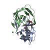
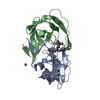
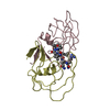
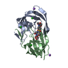
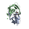
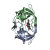
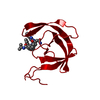
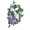
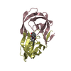
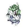
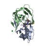
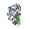
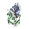
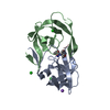
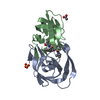
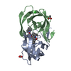
CRYSTALLOGRAPHIC ANALYSIS OF HUMAN IMMUNODEFICIENCY VIRUS 1 PROTEASE WITH AN ANALOG OF THE CONSERVED CA-P2 SUBSTRATE: INTERACTIONS WITH FREQUENTLY OCCURRING GLUTAMIC ACID RESIDUE AT P2' POSITION OF SUBSTRATES
Mass: 18.015 Da / Num. of mol.: 159 / Source method: isolated from a natural source / Formula: H2O
Nonpolymer details
PEPTIDE INHIBITOR 0Q4 HAS A REDUCED PEPTIDE (-CH2-NH) INSTEAD OF THE NORMAL PEPTIDE LINK (-CO-NH).
Sequence details
MUTATIONS Q7K, L33I, L63I HAVE BEEN MADE TO STABILIZE THE PROTEASE FROM AUTOPROTEOLYSIS, WHILE ...MUTATIONS Q7K, L33I, L63I HAVE BEEN MADE TO STABILIZE THE PROTEASE FROM AUTOPROTEOLYSIS, WHILE RETAINING ACTIVITY SIMILAR TO WILD-TYPE HIV-1 PROTEASE
-
Experimental details
-
Experiment
Experiment
Method: X-RAY DIFFRACTION / Number of used crystals: 1
-
Sample preparation
Crystal
Density Matthews: 2.14 Å3/Da / Density % sol: 42.41 % Description: THE DATA WAS COLLECTED ON AN R-AXIS IMAGING PLATE DETECTOR MOUNTED ON AN RU200 RIGAKU ROTATING ANODE X-RAY GENERATOR THAT WAS OPERATED AT 50 KV AND 100 MA WITH A 0.5-MM COLLIMATOR. EACH ...Description: THE DATA WAS COLLECTED ON AN R-AXIS IMAGING PLATE DETECTOR MOUNTED ON AN RU200 RIGAKU ROTATING ANODE X-RAY GENERATOR THAT WAS OPERATED AT 50 KV AND 100 MA WITH A 0.5-MM COLLIMATOR. EACH OSCILLATION FRAME WAS EXPOSED FOR 45 MIN. AT ROOM TEMPERATURE WITH 3.0 OSCILLATIONS. THE DISTANCE TO THE DETECTOR PLATE WAS SET AT 100 MM.
Crystal grow
pH: 4.7 / Details: pH 4.7
Crystal grow
*PLUS
Method: vapor diffusion / Details: macroseeding with 40-41% ammonium sulfate
In the structure databanks used in Yorodumi, some data are registered as the other names, "COVID-19 virus" and "2019-nCoV". Here are the details of the virus and the list of structure data.
Jan 31, 2019. EMDB accession codes are about to change! (news from PDBe EMDB page)
EMDB accession codes are about to change! (news from PDBe EMDB page)
The allocation of 4 digits for EMDB accession codes will soon come to an end. Whilst these codes will remain in use, new EMDB accession codes will include an additional digit and will expand incrementally as the available range of codes is exhausted. The current 4-digit format prefixed with “EMD-” (i.e. EMD-XXXX) will advance to a 5-digit format (i.e. EMD-XXXXX), and so on. It is currently estimated that the 4-digit codes will be depleted around Spring 2019, at which point the 5-digit format will come into force.
The EM Navigator/Yorodumi systems omit the EMD- prefix.
Related info.:Q: What is EMD? / ID/Accession-code notation in Yorodumi/EM Navigator
Yorodumi is a browser for structure data from EMDB, PDB, SASBDB, etc.
This page is also the successor to EM Navigator detail page, and also detail information page/front-end page for Omokage search.
The word "yorodu" (or yorozu) is an old Japanese word meaning "ten thousand". "mi" (miru) is to see.
Related info.:EMDB / PDB / SASBDB / Comparison of 3 databanks / Yorodumi Search / Aug 31, 2016. New EM Navigator & Yorodumi / Yorodumi Papers / Jmol/JSmol / Function and homology information / Changes in new EM Navigator and Yorodumi
 Movie
Movie Controller

 Yorodumi
 Open data
 Basic information
 Components
 Keywords
 Function and homology information
 Human immunodeficiency virus 1
 X-RAY DIFFRACTION /
  Authors
 Citation
 Structure visualization
 Downloads & links
 Other downloads


 PDBj
PDBj


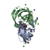
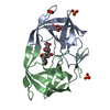
 Assembly
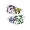
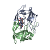
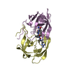

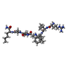
 Type: peptide-like, Peptide-like / Class: Inhibitor / Mass: 833.053 Da / Num. of mol.: 2 / Source method: obtained synthetically / Formula: C40H70N11O8 / Details: ANALOG OF THE CONSERVED CA-P2 SUBSTRATE
 Mass: 18.015 Da / Num. of mol.: 159 / Source method: isolated from a natural source / Formula: H2O
 Sample preparation
 Processing