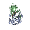
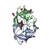
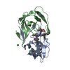
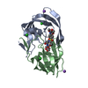
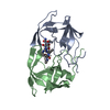
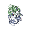
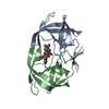
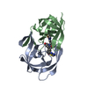
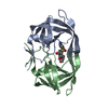
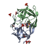
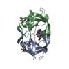
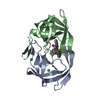
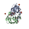
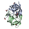
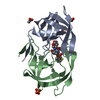
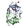
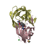
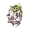
[English] 日本語
Yorodumi
- PDB-5uov: HIV-1 wild Type protease with GRL-1118A , an isophthalamide-deriv... -

+
Open data


ID or keywords:

Loading...

-
Basic information

Entry
Database: PDB / ID: 5uov
TitleHIV-1 wild Type protease with GRL-1118A , an isophthalamide-derived P2-P3 ligand with the sulfonamide isostere as the P2' group
ComponentsProtease
Keywordshydrolase/hydrolase inhibitor / an isophthalamide-derived P2-P3 ligand / HIV-1 protease inhibitor GRL-1118A / darunavir / multidrug-resistant / hydrolase inhibitor complex / hydrolase-hydrolase inhibitor complex
Function / homology
Function and homology information


HIV-1 retropepsin / : / retroviral ribonuclease H / exoribonuclease H / : / exoribonuclease H activity / host multivesicular body / DNA integration / RNA-directed DNA polymerase / viral genome integration into host DNA ...HIV-1 retropepsin / : / retroviral ribonuclease H / exoribonuclease H / : / exoribonuclease H activity / host multivesicular body / DNA integration / RNA-directed DNA polymerase / viral genome integration into host DNA / viral penetration into host nucleus / establishment of integrated proviral latency / RNA-directed DNA polymerase activity / Transferases; Transferring phosphorus-containing groups; Nucleotidyltransferases / RNA-DNA hybrid ribonuclease activity / viral nucleocapsid / DNA recombination / Hydrolases; Acting on ester bonds / DNA-directed DNA polymerase / aspartic-type endopeptidase activity / DNA-directed DNA polymerase activity / symbiont entry into host cell / symbiont-mediated suppression of host gene expression / lipid binding / host cell nucleus / host cell plasma membrane / virion membrane / structural molecule activity / proteolysis / DNA binding / RNA binding / zinc ion binding / membrane
Similarity search - Function
Reverse transcriptase connection / Reverse transcriptase connection domain / Reverse transcriptase thumb / Reverse transcriptase thumb domain / Integrase Zinc binding domain / Zinc finger integrase-type profile. / Integrase-like, N-terminal / Integrase DNA binding domain / Integrase, C-terminal domain superfamily, retroviral / Integrase, N-terminal zinc-binding domain ...Reverse transcriptase connection / Reverse transcriptase connection domain / Reverse transcriptase thumb / Reverse transcriptase thumb domain / Integrase Zinc binding domain / Zinc finger integrase-type profile. / Integrase-like, N-terminal / Integrase DNA binding domain / Integrase, C-terminal domain superfamily, retroviral / Integrase, N-terminal zinc-binding domain / Integrase, C-terminal, retroviral / Integrase DNA binding domain profile. / Immunodeficiency lentiviral matrix, N-terminal / gag gene protein p17 (matrix protein) / RNase H / Integrase core domain / Integrase, catalytic core / Integrase catalytic domain profile. / Retroviral nucleocapsid Gag protein p24, C-terminal domain / Gag protein p24 C-terminal domain / Retropepsin-like catalytic domain / Matrix protein, lentiviral and alpha-retroviral, N-terminal / Ribonuclease H domain / RNase H type-1 domain profile. / Reverse transcriptase (RNA-dependent DNA polymerase) / Reverse transcriptase domain / Reverse transcriptase (RT) catalytic domain profile. / Retropepsins / Retroviral aspartyl protease / Aspartyl protease, retroviral-type family profile. / Peptidase A2A, retrovirus, catalytic / Retrovirus capsid, C-terminal / Retroviral matrix protein / Retrovirus capsid, N-terminal / zinc finger / Zinc knuckle / Zinc finger, CCHC-type superfamily / Cathepsin D, subunit A; domain 1 / Acid Proteases / Zinc finger, CCHC-type / Zinc finger CCHC-type profile. / Ribonuclease H superfamily / Aspartic peptidase, active site / Eukaryotic and viral aspartyl proteases active site. / Aspartic peptidase domain superfamily / Ribonuclease H-like superfamily / Reverse transcriptase/Diguanylate cyclase domain / DNA/RNA polymerase superfamily / Beta Barrel / Mainly Beta
Similarity search - Domain/homology
Chem-8FP / ACETATE ION / Protease / Gag-Pol polyprotein
Similarity search - Component
Biological speciesHuman immunodeficiency virus 1
MethodX-RAY DIFFRACTION / SYNCHROTRON / MOLECULAR REPLACEMENT / Resolution: 1.33 Å
AuthorsWang, Y.-F. / Agniswamy, J. / Weber, I.T.
Funding support United States, Japan, 8items
OrganizationGrant numberCountry
National Institutes of Health/National Institute of General Medical Sciences (NIH/NIGMS)GM53386 United States
National Institutes of Health/National Institute of General Medical Sciences (NIH/NIGMS)GM62920 United States
Department of Energy (DOE, United States)W-31-109-Eng-38 United States
National Institutes of Health/National Cancer Institute (NIH/NCI)the Intramural Research Program of the Center for Cancer Research United States
Ministry of Education, Culture, Sports, Science and Technology (Japan)a Grant-in-Aid for Scientific Research (Priority Areas) Japan
Ministry of Health, Welfare, and Labora Grant for Promotion of AIDS Research Japan
Ministry of Education, Culture, Sports, Science and Technology (Japan)the Grant to the Cooperative Research Project on Clinical and Epidemiological Studies of Emerging and Reemerging Infectious Diseases (Renkei Jigyo) Japan
Purdue Universitythe Purdue University Center for Cancer Research United States
CitationJournal: Bioorg. Med. Chem. / Year: 2017
Title: Design of novel HIV-1 protease inhibitors incorporating isophthalamide-derived P2-P3 ligands: Synthesis, biological evaluation and X-ray structural studies of inhibitor-HIV-1 protease complex.
Authors: Ghosh, A.K. / Brindisi, M. / Nyalapatla, P.R. / Takayama, J. / Ella-Menye, J.R. / Yashchuk, S. / Agniswamy, J. / Wang, Y.F. / Aoki, M. / Amano, M. / Weber, I.T. / Mitsuya, H.
History
DepositionFeb 1, 2017Deposition site: RCSB / Processing site: RCSB
Revision 1.0May 10, 2017Provider: repository / Type: Initial release
Revision 1.1Sep 27, 2017Group: Author supporting evidence / Category: pdbx_audit_support / Item: _pdbx_audit_support.funding_organization
Revision 1.2Oct 4, 2017Group: Database references / Category: citation
Item: _citation.journal_volume / _citation.page_first / _citation.page_last
Revision 1.3Dec 4, 2019Group: Author supporting evidence / Category: pdbx_audit_support / Item: _pdbx_audit_support.funding_organization
Revision 1.4Oct 4, 2023Group: Data collection / Database references ...Data collection / Database references / Derived calculations / Refinement description
Category: chem_comp_atom / chem_comp_bond ...chem_comp_atom / chem_comp_bond / database_2 / pdbx_initial_refinement_model / pdbx_struct_conn_angle / struct_conn
Item: _database_2.pdbx_DOI / _database_2.pdbx_database_accession ..._database_2.pdbx_DOI / _database_2.pdbx_database_accession / _pdbx_struct_conn_angle.ptnr1_auth_seq_id / _pdbx_struct_conn_angle.ptnr3_auth_seq_id / _pdbx_struct_conn_angle.value / _struct_conn.pdbx_dist_value / _struct_conn.ptnr2_auth_seq_id

-
Structure visualization

Structure viewerMolecule:
MolmilJmol/JSmol

Downloads & links

-
Assembly

Deposited unit
A: Protease
B: Protease
hetero molecules


Theoretical massNumber of molelcules
Total (without water)22,4679
Polymers21,4812
Non-polymers9857
Water3,099172
1


  • Idetical with deposited unit
  • defined by author&software
TypeNameSymmetry operationNumber
identity operation1_555x,y,z1
Buried area5080 Å2
ΔGint-59 kcal/mol
Surface area9230 Å2
MethodPISA
Unit cell
Length a, b, c (Å)58.179, 86.515, 45.945
Angle α, β, γ (deg.)90.00, 90.00, 90.00
Int Tables number18
Space group name H-MP21212

-
Components

-
Protein , 1 types, 2 molecules AB

#1: Protein Protease /


Mass: 10740.677 Da / Num. of mol.: 2 / Mutation: Q7K, L33I, L63I, C67A, C95A
Source method: isolated from a genetically manipulated source
Source: (gene. exp.) Human immunodeficiency virus 1 / Gene: pol / Plasmid: pET11a / Production host: Escherichia coli (E. coli) / Strain (production host): Bl21 (de3) / References: UniProt: C8B467, UniProt: P03366*PLUS

-
Non-polymers , 6 types, 179 molecules

#2: Chemical ChemComp-8FP / N-[(2S,3R)-3-hydroxy-4-{[(4-methoxyphenyl)sulfonyl](2-methylpropyl)amino}-1-phenylbutan-2-yl]-3-[(2R)-2-(4-methyl-1,3-thiazol-2-yl)pyrrolidine-1-carbonyl]benzamide


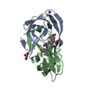
Mass: 704.898 Da / Num. of mol.: 1 / Source method: obtained synthetically / Formula: C37H44N4O6S2
#3: Chemical ChemComp-NA / SODIUM ION


Mass: 22.990 Da / Num. of mol.: 1 / Source method: obtained synthetically / Formula: Na
#4: Chemical ChemComp-CL / CHLORIDE ION / Chloride


Mass: 35.453 Da / Num. of mol.: 3 / Source method: obtained synthetically / Formula: Cl
#5: Chemical ChemComp-GOL / GLYCEROL / GLYCERIN / PROPANE-1,2,3-TRIOL / Glycerol


Mass: 92.094 Da / Num. of mol.: 1 / Source method: obtained synthetically / Formula: C3H8O3
#6: Chemical ChemComp-ACT / ACETATE ION / Acetate


Mass: 59.044 Da / Num. of mol.: 1 / Source method: obtained synthetically / Formula: C2H3O2
#7: Water ChemComp-HOH / water / Water


Mass: 18.015 Da / Num. of mol.: 172 / Source method: isolated from a natural source / Formula: H2O

-
Experimental details

-
Experiment

ExperimentMethod: X-RAY DIFFRACTION / Number of used crystals: 1

-
Sample preparation

CrystalDensity Matthews: 2.69 Å3/Da / Density % sol: 54.28 %
Crystal growTemperature: 298 K / Method: vapor diffusion, hanging drop / pH: 4.8 / Details: 1.28 M NaCl, 0.1 M Sodium Acetate, pH 4.8 / PH range: 4.8

-
Data collection

DiffractionMean temperature: 100 K
Diffraction sourceSource: SYNCHROTRON / Site: APS / Beamline: 22-BM / Wavelength: 1 Å
DetectorType: MARMOSAIC 225 mm CCD / Detector: CCD / Date: Nov 20, 2008
RadiationProtocol: SINGLE WAVELENGTH / Monochromatic (M) / Laue (L): M / Scattering type: x-ray
Radiation wavelengthWavelength: 1 Å / Relative weight: 1
ReflectionResolution: 1.33→50 Å / Num. obs: 52049 / % possible obs: 96.2 % / Redundancy: 6.5 % / Rmerge(I) obs: 0.072 / Net I/σ(I): 18.2
Reflection shellResolution: 1.33→1.38 Å / Redundancy: 2.2 % / Rmerge(I) obs: 0.456 / Mean I/σ(I) obs: 2.2 / % possible all: 72.4

-
Processing

Software
NameClassification
SHELXL-97refinement
HKL-2000data reduction
HKL-2000data scaling
AMoREphasing
RefinementMethod to determine structure: MOLECULAR REPLACEMENT
Starting model: 3DJK
Resolution: 1.33→50 Å / Num. parameters: 16582 / Num. restraintsaints: 21576 / Cross valid method: FREE R / σ(F): 0 / Stereochemistry target values: ENGH AND HUBER
Details: ANISOTROPIC REFINEMENT REDUCED FREE R (NO CUTOFF) BY ?
RfactorNum. reflection% reflectionSelection details
Rfree0.1834 2602 5 %RANDOM
Rwork0.1524 ---
all0.154 ---
obs-51979 96.2 %-
Refine analyzeNum. disordered residues: 38 / Occupancy sum hydrogen: 0 / Occupancy sum non hydrogen: 1706.85
Refinement stepCycle: LAST / Resolution: 1.33→50 Å
ProteinNucleic acidLigandSolventTotal
Num. atoms1512 0 49 182 1743
Refine LS restraints
Refine-IDTypeDev ideal
X-RAY DIFFRACTIONs_bond_d0.012
X-RAY DIFFRACTIONs_angle_d0.029
X-RAY DIFFRACTIONs_similar_dist0
X-RAY DIFFRACTIONs_from_restr_planes0.0298
X-RAY DIFFRACTIONs_zero_chiral_vol0.066
X-RAY DIFFRACTIONs_non_zero_chiral_vol0.08
X-RAY DIFFRACTIONs_anti_bump_dis_restr0.018
X-RAY DIFFRACTIONs_rigid_bond_adp_cmpnt0.004
X-RAY DIFFRACTIONs_similar_adp_cmpnt0.056
X-RAY DIFFRACTIONs_approx_iso_adps0.082

+
About Yorodumi

-
News

-
Feb 9, 2022. New format data for meta-information of EMDB entries

New format data for meta-information of EMDB entries

  • Version 3 of the EMDB header file is now the official format.
  • The previous official version 1.9 will be removed from the archive.

Related info.:EMDB header

External links:wwPDB to switch to version 3 of the EMDB data model

-
Aug 12, 2020. Covid-19 info

Covid-19 info

URL: https://pdbj.org/emnavi/covid19.php

New page: Covid-19 featured information page in EM Navigator.

Related info.:Covid-19 info / Mar 5, 2020. Novel coronavirus structure data

+
Mar 5, 2020. Novel coronavirus structure data

Novel coronavirus structure data

Related info.:Yorodumi Speices / Aug 12, 2020. Covid-19 info

External links:COVID-19 featured content - PDBj / Molecule of the Month (242):Coronavirus Proteases

+
Jan 31, 2019. EMDB accession codes are about to change! (news from PDBe EMDB page)

EMDB accession codes are about to change! (news from PDBe EMDB page)

  • The allocation of 4 digits for EMDB accession codes will soon come to an end. Whilst these codes will remain in use, new EMDB accession codes will include an additional digit and will expand incrementally as the available range of codes is exhausted. The current 4-digit format prefixed with “EMD-” (i.e. EMD-XXXX) will advance to a 5-digit format (i.e. EMD-XXXXX), and so on. It is currently estimated that the 4-digit codes will be depleted around Spring 2019, at which point the 5-digit format will come into force.
  • The EM Navigator/Yorodumi systems omit the EMD- prefix.

Related info.:Q: What is EMD? / ID/Accession-code notation in Yorodumi/EM Navigator

External links:EMDB Accession Codes are Changing Soon! / Contact to PDBj

+
Jul 12, 2017. Major update of PDB

Major update of PDB

  • wwPDB released updated PDB data conforming to the new PDBx/mmCIF dictionary.
  • This is a major update changing the version number from 4 to 5, and with Remediation, in which all the entries are updated.
  • In this update, many items about electron microscopy experimental information are reorganized (e.g. em_software).
  • Now, EM Navigator and Yorodumi are based on the updated data.

External links:wwPDB Remediation / Enriched Model Files Conforming to OneDep Data Standards Now Available in the PDB FTP Archive

-
Yorodumi

Thousand views of thousand structures

  • Yorodumi is a browser for structure data from EMDB, PDB, SASBDB, etc.
  • This page is also the successor to EM Navigator detail page, and also detail information page/front-end page for Omokage search.
  • The word "yorodu" (or yorozu) is an old Japanese word meaning "ten thousand". "mi" (miru) is to see.

Related info.:EMDB / PDB / SASBDB / Comparison of 3 databanks / Yorodumi Search / Aug 31, 2016. New EM Navigator & Yorodumi / Yorodumi Papers / Jmol/JSmol / Function and homology information / Changes in new EM Navigator and Yorodumi

Read more