 Movie
Movie Controller

[English] 日本語
 Yorodumi
- PDB-6pu8: Room temperature X-ray structure of HIV-1 protease triple mutant ... -
+ Open data

- Basic information
| Entry | Database: PDB / ID: 6pu8 | ||||||
|---|---|---|---|---|---|---|---|
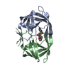
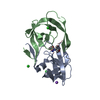
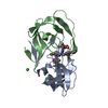
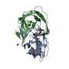
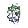
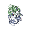
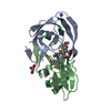
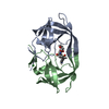
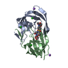
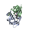
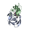
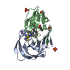
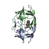
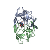
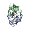
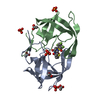
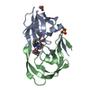
| Title | Room temperature X-ray structure of HIV-1 protease triple mutant (V32I,I47V,V82I) with tetrahedral intermediate of keto-darunavir | ||||||
 Components Components | HIV-1 protease | ||||||
 Keywords Keywords | HYDROLASE / HIV-1 protease / aspartic protease / tetrahedral intermediate inhibitor / homodimer | ||||||
| Function / homology |  Function and homology information Function and homology informationhost multivesicular body / aspartic-type endopeptidase activity / virion membrane / proteolysis / identical protein binding Similarity search - Function | ||||||
| Biological species |   Human immunodeficiency virus 1 Human immunodeficiency virus 1 | ||||||
| Method |  X-RAY DIFFRACTION / MOLECULAR REPLACEMENT / Resolution: 1.8 Å X-RAY DIFFRACTION / MOLECULAR REPLACEMENT / Resolution: 1.8 Å | ||||||
 Authors Authors | Kovalevsky, A. / Das, A. | ||||||
 Citation Citation | Journal: Acs Omega / Year: 2020 Title: Visualizing Tetrahedral Oxyanion Bound in HIV-1 Protease Using Neutrons: Implications for the Catalytic Mechanism and Drug Design. Authors: Kumar, M. / Mandal, K. / Blakeley, M.P. / Wymore, T. / Kent, S.B.H. / Louis, J.M. / Das, A. / Kovalevsky, A. | ||||||
| History |
|
- Structure visualization
| Structure viewer | Molecule: MolmilJmol/JSmol |
|---|
- Downloads & links
-Download
| PDBx/mmCIF format | 6pu8.cif.gz | 55.9 KB | Display | PDBx/mmCIF format |
|---|---|---|---|---|
| PDB format | pdb6pu8.ent.gz | 38.2 KB | Display | PDB format |
| PDBx/mmJSON format | 6pu8.json.gz | Tree view | PDBx/mmJSON format | |
| Others |  Other downloads Other downloads |
-Validation report
| Arichive directory | https://data.pdbj.org/pub/pdb/validation_reports/pu/6pu8ftp://data.pdbj.org/pub/pdb/validation_reports/pu/6pu8 | HTTPS FTP |
|---|
-Related structure data
| Related structure data |  6kmpC 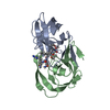 6ptpC 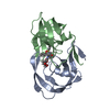 5e5jS S: Starting model for refinement C: citing same article ( |
|---|---|
| Similar structure data |


-Links
 PDBj
PDBj
- Assembly
| Deposited unit | 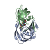
| ||||||||
|---|---|---|---|---|---|---|---|---|---|
| 1 |
| ||||||||
| Unit cell |
|
-Components
| #1: Protein | Mass: 10754.703 Da / Num. of mol.: 2 / Mutation: V32I,I47V,V82I Source method: isolated from a genetically manipulated source Source: (gene. exp.) Human immunodeficiency virus 1 / Gene: pol / Production host:  #2: Chemical | ChemComp-P3V / ( |   Mass: 563.663 Da / Num. of mol.: 1 / Source method: obtained synthetically / Formula: C27H37N3O8S Mass: 563.663 Da / Num. of mol.: 1 / Source method: obtained synthetically / Formula: C27H37N3O8S#3: Water | ChemComp-HOH / |  Mass: 18.015 Da / Num. of mol.: 121 / Source method: isolated from a natural source / Formula: H2O Mass: 18.015 Da / Num. of mol.: 121 / Source method: isolated from a natural source / Formula: H2OHas ligand of interest | Y | |
|---|
-Experimental details
-Experiment
| Experiment | Method: X-RAY DIFFRACTION / Number of used crystals: 1 |
|---|
- Sample preparation
| Crystal | Density Matthews: 2.81 Å3/Da / Density % sol: 56.28 % |
|---|---|
| Crystal grow | Temperature: 293 K / Method: vapor diffusion, sitting drop / pH: 6 / Details: 0.1 M MES, 1.0 M NaCl, pH 6.0 |
-Data collection
| Diffraction | Mean temperature: 293 K / Serial crystal experiment: N |
|---|---|
| Diffraction source | Source: ROTATING ANODE / Type: RIGAKU MICROMAX-007 / Wavelength: 1.5406 Å |
| Detector | Type: RIGAKU RAXIS IV++ / Detector: IMAGE PLATE / Date: Nov 21, 2016 |
| Radiation | Protocol: SINGLE WAVELENGTH / Monochromatic (M) / Laue (L): M / Scattering type: x-ray |
| Radiation wavelength | Wavelength: 1.5406 Å / Relative weight: 1 |
| Reflection | Resolution: 1.8→40 Å / Num. obs: 22743 / % possible obs: 98 % / Redundancy: 3.9 % / Rmerge(I) obs: 0.037 / Net I/σ(I): 29.4 |
| Reflection shell | Resolution: 1.8→1.86 Å / Redundancy: 3.8 % / Rmerge(I) obs: 0.481 / Mean I/σ(I) obs: 2.2 / Num. unique obs: 2253 / % possible all: 99 |
- Processing
| Software |
| |||||||||||||||||||||||||||||||||
|---|---|---|---|---|---|---|---|---|---|---|---|---|---|---|---|---|---|---|---|---|---|---|---|---|---|---|---|---|---|---|---|---|---|---|
| Refinement | Method to determine structure: MOLECULAR REPLACEMENT Starting model: 5E5J Resolution: 1.8→20 Å / Num. parameters: 6699 / Num. restraintsaints: 6296 / Cross valid method: FREE R-VALUE / σ(F): 0 / Stereochemistry target values: ENGH AND HUBER
| |||||||||||||||||||||||||||||||||
| Refine analyze | Num. disordered residues: 0 / Occupancy sum hydrogen: 0 / Occupancy sum non hydrogen: 1658.5 | |||||||||||||||||||||||||||||||||
| Refinement step | Cycle: 1 / Resolution: 1.8→20 Å
| |||||||||||||||||||||||||||||||||
| Refine LS restraints |
|
