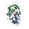
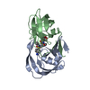
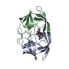
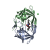
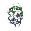
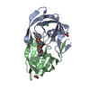
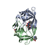
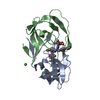
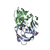
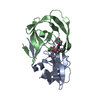
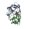
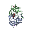
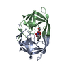
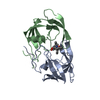
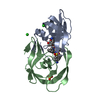
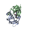
Entry Database : PDB / ID : 4jecTitle Joint neutron and X-ray structure of per-deuterated HIV-1 protease in complex with clinical inhibitor amprenavir HIV-1 protease Keywords / / / / Function / homology Function Domain/homology Component
/ / / / / / / / / / / / / / / / / / / / / / / / / / / / / / / / / / / / / / / / / / / / / / / / / / / / / / / / / / / / / / / / / / / / / / / / / / / / / / / / / / / / / / / / / / / / / / / / / / / Biological species Method / / / / Resolution : 2.01 Å Authors Kovalevsky, A.Y. / Weber, I.T. / Langan, P. Journal : J.Med.Chem. / Year : 2013Title : Joint X-ray/Neutron Crystallographic Study of HIV-1 Protease with Clinical Inhibitor Amprenavir: Insights for Drug Design.Authors : Weber, I.T. / Waltman, M.J. / Mustyakimov, M. / Blakeley, M.P. / Keen, D.A. / Ghosh, A.K. / Langan, P. / Kovalevsky, A.Y. History Deposition Feb 26, 2013 Deposition site / Processing site Revision 1.0 Jul 24, 2013 Provider / Type Revision 1.1 Jun 13, 2018 Group / Refinement descriptionCategory diffrn_detector / diffrn_radiation ... diffrn_detector / diffrn_radiation / diffrn_radiation_wavelength / diffrn_source / refine Item _diffrn_detector.details / _diffrn_detector.pdbx_collection_date ... _diffrn_detector.details / _diffrn_detector.pdbx_collection_date / _diffrn_detector.type / _diffrn_radiation.pdbx_diffrn_protocol / _diffrn_radiation.pdbx_monochromatic_or_laue_m_l / _diffrn_radiation.pdbx_scattering_type / _diffrn_radiation_wavelength.wavelength / _diffrn_source.pdbx_synchrotron_beamline / _diffrn_source.pdbx_wavelength / _diffrn_source.pdbx_wavelength_list / _diffrn_source.source / _diffrn_source.type / _refine.ls_R_factor_R_free / _refine.ls_R_factor_R_work / _refine.ls_R_factor_obs / _refine.ls_d_res_high / _refine.ls_d_res_low / _refine.ls_number_reflns_R_free / _refine.ls_number_reflns_all / _refine.ls_number_reflns_obs / _refine.ls_percent_reflns_R_free / _refine.ls_percent_reflns_obs / _refine.pdbx_diffrn_id / _refine.pdbx_ls_sigma_F / _refine.pdbx_refine_id Revision 1.2 Feb 28, 2024 Group Data collection / Database references ... Data collection / Database references / Derived calculations / Refinement description Category chem_comp_atom / chem_comp_bond ... chem_comp_atom / chem_comp_bond / database_2 / refine_hist / struct_ref_seq_dif / struct_site Item _database_2.pdbx_DOI / _database_2.pdbx_database_accession ... _database_2.pdbx_DOI / _database_2.pdbx_database_accession / _refine_hist.d_res_high / _refine_hist.d_res_low / _struct_ref_seq_dif.details / _struct_site.pdbx_auth_asym_id / _struct_site.pdbx_auth_comp_id / _struct_site.pdbx_auth_seq_id Revision 1.3 Nov 12, 2025 Group / Structure summary / Category / pdbx_entry_details / Item
Show all Show less
 Movie
Movie Controller

 Yorodumi
 Open data
 Basic information
 Components
 Keywords
 Function and homology information
 Human immunodeficiency virus type 1
 X-RAY DIFFRACTION / NUCLEAR REACTOR /
  Authors
 Citation
 Structure visualization
 Downloads & links
 Other downloads
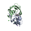


 PDBj
PDBj


 Assembly
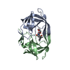


 Mass: 35.453 Da / Num. of mol.: 1 / Source method: obtained synthetically / Formula: Cl
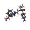
 Mass: 505.627 Da / Num. of mol.: 1 / Source method: obtained synthetically / Formula: C25H35N3O6S / Comment: medication*YM
 Mass: 18.015 Da / Num. of mol.: 131 / Source method: isolated from a natural source / Formula: D2O
 Sample preparation
 Processing