integrase activity / Integration of viral DNA into host genomic DNA / Autointegration results in viral DNA circles / Minus-strand DNA synthesis / Plus-strand DNA synthesis / Uncoating of the HIV Virion / 2-LTR circle formation / Vpr-mediated nuclear import of PICs / Early Phase of HIV Life Cycle / Integration of provirus ...integrase activity / Integration of viral DNA into host genomic DNA / Autointegration results in viral DNA circles / Minus-strand DNA synthesis / Plus-strand DNA synthesis / Uncoating of the HIV Virion / 2-LTR circle formation / Vpr-mediated nuclear import of PICs / Early Phase of HIV Life Cycle / Integration of provirus / APOBEC3G mediated resistance to HIV-1 infection / Binding and entry of HIV virion / viral life cycle / HIV-1 retropepsin / symbiont-mediated activation of host apoptosis / retroviral ribonuclease H / exoribonuclease H / exoribonuclease H activity / Assembly Of The HIV Virion / protein processing / viral genome integration into host DNA / Budding and maturation of HIV virion / establishment of integrated proviral latency / RNA-directed DNA polymerase / RNA stem-loop binding / viral penetration into host nucleus / host multivesicular body / RNA-directed DNA polymerase activity / RNA-DNA hybrid ribonuclease activity / Transferases; Transferring phosphorus-containing groups; Nucleotidyltransferases / peptidase activity / host cell / viral nucleocapsid / DNA recombination / DNA-directed DNA polymerase / aspartic-type endopeptidase activity / Hydrolases; Acting on ester bonds / DNA-directed DNA polymerase activity / symbiont-mediated suppression of host gene expression / viral translational frameshifting / symbiont entry into host cell / lipid binding / host cell nucleus / host cell plasma membrane / virion membrane / structural molecule activity / DNA binding / zinc ion binding / identical protein binding Similarity search - Function
1: THERE ARE OXYGENS COVALENTLY BOUND TO SG CYS A 67 AND SG CYS B 67. THEY ARE PRESENTED AS HETATM RECORDS FOLLOWING THE END OF EACH CHAIN.
-
Components
#1: Protein
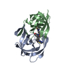
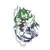
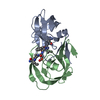
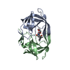
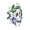
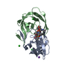
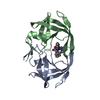
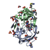
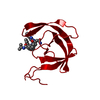
HIV-1PROTEASE
Mass: 10805.729 Da / Num. of mol.: 2 Source method: isolated from a genetically manipulated source Source: (gene. exp.) Human immunodeficiency virus 1 / Production host: Escherichia coli (E. coli) / References: UniProt: P04585
Mass: 18.015 Da / Num. of mol.: 90 / Source method: isolated from a natural source / Formula: H2O
Has protein modification
Y
Nonpolymer details
THE SUBCOMPONENT CAV IN INHIBITOR 1ZK IS THE DIPEPTIDE ISOSTERE CYCLOHEXYL ALANINE PSI(CHOH-CHOH) ...THE SUBCOMPONENT CAV IN INHIBITOR 1ZK IS THE DIPEPTIDE ISOSTERE CYCLOHEXYL ALANINE PSI(CHOH-CHOH)VALINE. THE VALINE HAS A C-OH GROUP IN PLACE OF THE USUAL MAIN CHAIN N ATOM.
-
Experimental details
-
Experiment
Experiment
Method: X-RAY DIFFRACTION
-
Sample preparation
Crystal
Density Matthews: 2.1 Å3/Da / Density % sol: 41.56 %
Crystal grow
*PLUS
pH: 6.8 / Method: vapor diffusion, hanging drop
Components of the solutions
*PLUS
ID
Conc.
Common name
Crystal-ID
Sol-ID
1
20 %(v/v)
ammoniumsulfate
1
reservoir
2
0.1M
sodiumacetate
1
reservoir
3
10mg/ml
proteincomplex
1
drop
-
Data collection
Radiation
Scattering type: x-ray
Radiation wavelength
Relative weight: 1
-
Processing
Software
Name: PROLSQ / Classification: refinement
Refinement
Rfactor obs: 0.169 / Highest resolution: 2 Å
Refinement step
Cycle: LAST / Highest resolution: 2 Å
Protein
Nucleic acid
Ligand
Solvent
Total
Num. atoms
1516
0
59
90
1665
Refine LS restraints
Refine-ID
Type
Dev ideal
Dev ideal target
X-RAY DIFFRACTION
p_bond_d
0.02
0.02
X-RAY DIFFRACTION
p_angle_d
0.049
0.035
X-RAY DIFFRACTION
p_angle_deg
X-RAY DIFFRACTION
p_planar_d
0.056
0.05
X-RAY DIFFRACTION
p_hb_or_metal_coord
X-RAY DIFFRACTION
p_mcbond_it
1.475
1.5
X-RAY DIFFRACTION
p_mcangle_it
2.21
2
X-RAY DIFFRACTION
p_scbond_it
4.215
3
X-RAY DIFFRACTION
p_scangle_it
5.934
4
X-RAY DIFFRACTION
p_plane_restr
0.017
0.02
X-RAY DIFFRACTION
p_chiral_restr
0.201
0.15
X-RAY DIFFRACTION
p_singtor_nbd
0.191
0.3
X-RAY DIFFRACTION
p_multtor_nbd
0.201
0.3
X-RAY DIFFRACTION
p_xhyhbond_nbd
0.185
0.3
X-RAY DIFFRACTION
p_xyhbond_nbd
X-RAY DIFFRACTION
p_planar_tor
2.8
3
X-RAY DIFFRACTION
p_staggered_tor
17.6
10
X-RAY DIFFRACTION
p_orthonormal_tor
17.2
20
X-RAY DIFFRACTION
p_transverse_tor
X-RAY DIFFRACTION
p_special_tor
Refinement
*PLUS
Highest resolution: 2 Å / Lowest resolution: 10 Å / Num. reflection obs: 10110 / Rfactor obs: 0.169
Solvent computation
*PLUS
Displacement parameters
*PLUS
+
About Yorodumi
-
News
-
Feb 9, 2022. New format data for meta-information of EMDB entries
New format data for meta-information of EMDB entries
Version 3 of the EMDB header file is now the official format.
The previous official version 1.9 will be removed from the archive.
In the structure databanks used in Yorodumi, some data are registered as the other names, "COVID-19 virus" and "2019-nCoV". Here are the details of the virus and the list of structure data.
Jan 31, 2019. EMDB accession codes are about to change! (news from PDBe EMDB page)
EMDB accession codes are about to change! (news from PDBe EMDB page)
The allocation of 4 digits for EMDB accession codes will soon come to an end. Whilst these codes will remain in use, new EMDB accession codes will include an additional digit and will expand incrementally as the available range of codes is exhausted. The current 4-digit format prefixed with “EMD-” (i.e. EMD-XXXX) will advance to a 5-digit format (i.e. EMD-XXXXX), and so on. It is currently estimated that the 4-digit codes will be depleted around Spring 2019, at which point the 5-digit format will come into force.
The EM Navigator/Yorodumi systems omit the EMD- prefix.
Related info.:Q: What is EMD? / ID/Accession-code notation in Yorodumi/EM Navigator
Yorodumi is a browser for structure data from EMDB, PDB, SASBDB, etc.
This page is also the successor to EM Navigator detail page, and also detail information page/front-end page for Omokage search.
The word "yorodu" (or yorozu) is an old Japanese word meaning "ten thousand". "mi" (miru) is to see.
Related info.:EMDB / PDB / SASBDB / Comparison of 3 databanks / Yorodumi Search / Aug 31, 2016. New EM Navigator & Yorodumi / Yorodumi Papers / Jmol/JSmol / Function and homology information / Changes in new EM Navigator and Yorodumi
 Movie
Movie Controller

 Yorodumi
 Open data
 Basic information
 Components
 Keywords
 Function and homology information
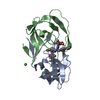
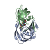
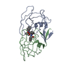
 Human immunodeficiency virus 1
 X-RAY DIFFRACTION / Resolution: 2 Å
  Authors
 Citation
 Structure visualization
 Downloads & links
 Other downloads
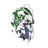


 PDBj
PDBj


 Assembly
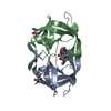

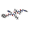
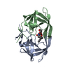
 Type: peptide-like, Peptide-like / Class: Inhibitor / Mass: 813.016 Da / Num. of mol.: 1 / Source method: obtained synthetically / Formula: C45H62N7O7 / References: Noa-His-Cha.psi.[CHOHCHOH]Val-Ile-Amp, U75875
 Mass: 18.015 Da / Num. of mol.: 90 / Source method: isolated from a natural source / Formula: H2O
 Sample preparation
 Processing