 Movie
Movie Controller

[English] 日本語
 Yorodumi
- PDB-1vij: HIV-1 PROTEASE COMPLEXED WITH THE INHIBITOR HOE/BAY 793 HEXAGONAL FORM -
+ Open data

- Basic information
| Entry | Database: PDB / ID: 1vij | ||||||
|---|---|---|---|---|---|---|---|
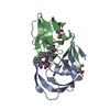
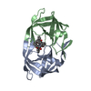
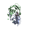
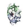
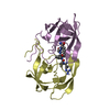
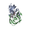
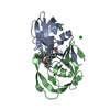
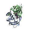
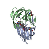
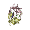
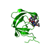
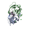
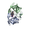
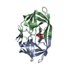
| Title | HIV-1 PROTEASE COMPLEXED WITH THE INHIBITOR HOE/BAY 793 HEXAGONAL FORM | ||||||
 Components Components | HIV-1 PROTEASE | ||||||
 Keywords Keywords | ASPARTYL PROTEASE / HUMAN IMMUNODEFICIENCY VIRUS / HOE/BAY 793: INHIBITOR DESIGN | ||||||
| Function / homology |  Function and homology information Function and homology informationHIV-1 retropepsin / symbiont-mediated activation of host apoptosis / retroviral ribonuclease H / exoribonuclease H / exoribonuclease H activity / DNA integration / viral genome integration into host DNA / establishment of integrated proviral latency / RNA-directed DNA polymerase / RNA stem-loop binding ...HIV-1 retropepsin / symbiont-mediated activation of host apoptosis / retroviral ribonuclease H / exoribonuclease H / exoribonuclease H activity / DNA integration / viral genome integration into host DNA / establishment of integrated proviral latency / RNA-directed DNA polymerase / RNA stem-loop binding / viral penetration into host nucleus / host multivesicular body / RNA-directed DNA polymerase activity / RNA-DNA hybrid ribonuclease activity / Transferases; Transferring phosphorus-containing groups; Nucleotidyltransferases / host cell / viral nucleocapsid / DNA recombination / DNA-directed DNA polymerase / aspartic-type endopeptidase activity / Hydrolases; Acting on ester bonds / DNA-directed DNA polymerase activity / symbiont-mediated suppression of host gene expression / viral translational frameshifting / symbiont entry into host cell / lipid binding / host cell nucleus / host cell plasma membrane / virion membrane / structural molecule activity / proteolysis / DNA binding / zinc ion binding Similarity search - Function | ||||||
| Biological species |   Human immunodeficiency virus 1 Human immunodeficiency virus 1 | ||||||
| Method |  X-RAY DIFFRACTION / Resolution: 2.4 Å X-RAY DIFFRACTION / Resolution: 2.4 Å | ||||||
 Authors Authors | Lange-Savage, G. / Berchtold, H. / Liesum, A. / Hilgenfeld, R. | ||||||
 Citation Citation | Journal: Eur.J.Biochem. / Year: 1997 Title: Structure of HOE/BAY 793 complexed to human immunodeficiency virus (HIV-1) protease in two different crystal forms--structure/function relationship and influence of crystal packing. Authors: Lange-Savage, G. / Berchtold, H. / Liesum, A. / Budt, K.H. / Peyman, A. / Knolle, J. / Sedlacek, J. / Fabry, M. / Hilgenfeld, R. #1: Journal: Eur.J.Biochem. / Year: 1997Title: Erratum. Structure of Hoe/Bay 793 Complexed to Human Immunodeficiency Virus (HIV-1) Protease in Two Different Crystal Forms--Structure/Function Relationship and Influence of Crystal Packing Authors: Lange-Savage, G. / Berchtold, H. / Liesum, A. / Budt, K.H. / Peyman, A. / Knolle, J. / Sedlacek, J. / Fabry, M. / Hilgenfeld, R. #2: Journal: Annu.Rev.Biochem. / Year: 1993Title: Structure-Based Inhibitors of HIV-1 Protease Authors: Wlodawer, A. / Erickson, J.W. | ||||||
| History |
|
- Structure visualization
| Structure viewer | Molecule: MolmilJmol/JSmol |
|---|
- Downloads & links
-Download
| PDBx/mmCIF format | 1vij.cif.gz | 55.4 KB | Display | PDBx/mmCIF format |
|---|---|---|---|---|
| PDB format | pdb1vij.ent.gz | 39.6 KB | Display | PDB format |
| PDBx/mmJSON format | 1vij.json.gz | Tree view | PDBx/mmJSON format | |
| Others |  Other downloads Other downloads |
-Validation report
| Arichive directory | https://data.pdbj.org/pub/pdb/validation_reports/vi/1vijftp://data.pdbj.org/pub/pdb/validation_reports/vi/1vij | HTTPS FTP |
|---|
-Related structure data


-Links
 PDBj
PDBj

- Assembly
| Deposited unit | 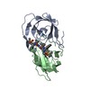
| ||||||||
|---|---|---|---|---|---|---|---|---|---|
| 1 |
| ||||||||
| Unit cell |
|
-Components
| #1: Protein | Mass: 10830.781 Da / Num. of mol.: 2 Source method: isolated from a genetically manipulated source Source: (gene. exp.) Human immunodeficiency virus 1 / Genus: Lentivirus / Production host:  #2: Chemical | ChemComp-BAY / |   Mass: 1131.487 Da / Num. of mol.: 1 / Source method: obtained synthetically / Formula: C64H82N4O10S2 Mass: 1131.487 Da / Num. of mol.: 1 / Source method: obtained synthetically / Formula: C64H82N4O10S2#3: Water | ChemComp-HOH / |  Mass: 18.015 Da / Num. of mol.: 101 / Source method: isolated from a natural source / Formula: H2O Mass: 18.015 Da / Num. of mol.: 101 / Source method: isolated from a natural source / Formula: H2O |
|---|
-Experimental details
-Experiment
| Experiment | Method: X-RAY DIFFRACTION |
|---|
- Sample preparation
| Crystal | Density Matthews: 2.28 Å3/Da / Density % sol: 46.08 % | ||||||||||||||||||||||||||||||
|---|---|---|---|---|---|---|---|---|---|---|---|---|---|---|---|---|---|---|---|---|---|---|---|---|---|---|---|---|---|---|---|
| Crystal | *PLUS Density % sol: 37 % | ||||||||||||||||||||||||||||||
| Crystal grow | *PLUS pH: 5.8 / Method: vapor diffusion, hanging drop | ||||||||||||||||||||||||||||||
| Components of the solutions | *PLUS
|
-Data collection
| Radiation | Scattering type: x-ray |
|---|---|
| Radiation wavelength | Relative weight: 1 |
| Reflection | *PLUS Highest resolution: 2.4 Å / Num. obs: 5468 / % possible obs: 90 % / Num. measured all: 18894 / Rmerge(I) obs: 0.07 |
| Reflection shell | *PLUS Highest resolution: 2.4 Å / Lowest resolution: 2.5 Å / % possible obs: 93 % |
- Processing
| Software | Name: PROLSQ / Classification: refinement | |||||||||||||||||||||||||||||||||||||||||||||||||||||||||||||||
|---|---|---|---|---|---|---|---|---|---|---|---|---|---|---|---|---|---|---|---|---|---|---|---|---|---|---|---|---|---|---|---|---|---|---|---|---|---|---|---|---|---|---|---|---|---|---|---|---|---|---|---|---|---|---|---|---|---|---|---|---|---|---|---|---|
| Refinement | Resolution: 2.4→8 Å / σ(F): 0 Details: RMS DEVIATIONS FROM IDEAL VALUES ANGLE DISTANCE (DEGREES) : 4.40
| |||||||||||||||||||||||||||||||||||||||||||||||||||||||||||||||
| Displacement parameters | Biso mean: 24 Å2 | |||||||||||||||||||||||||||||||||||||||||||||||||||||||||||||||
| Refinement step | Cycle: LAST / Resolution: 2.4→8 Å
| |||||||||||||||||||||||||||||||||||||||||||||||||||||||||||||||
| Refine LS restraints |
| |||||||||||||||||||||||||||||||||||||||||||||||||||||||||||||||
| Refine LS restraints | *PLUS Type: p_plane_restr / Dev ideal: 0.016 |
