 Movie
Movie Controller

[English] 日本語
 Yorodumi
- PDB-1hvu: HUMAN IMMUNODEFICIENCY VIRUS TYPE 1 REVERSE TRANSCRIPTASE COMPLEX... -
+ Open data

- Basic information
| Entry | Database: PDB / ID: 1hvu | ||||||
|---|---|---|---|---|---|---|---|
| Title | HUMAN IMMUNODEFICIENCY VIRUS TYPE 1 REVERSE TRANSCRIPTASE COMPLEXED WITH A 33-BASE NUCLEOTIDE RNA PSEUDOKNOT | ||||||
 Components Components |
| ||||||
 Keywords Keywords | TRANSFERASE/RNA / COMPLEX (NUCLEOTIDYLTRANSFERASE-RNA) / HYDROLASE / ASPARTYL PROTEASE / ENDONUCLEASE / TRANSFERASE-RNA complex | ||||||
| Function / homology |  Function and homology information Function and homology informationHIV-1 retropepsin / symbiont-mediated activation of host apoptosis / retroviral ribonuclease H / exoribonuclease H / exoribonuclease H activity / DNA integration / viral genome integration into host DNA / establishment of integrated proviral latency / RNA-directed DNA polymerase / RNA stem-loop binding ...HIV-1 retropepsin / symbiont-mediated activation of host apoptosis / retroviral ribonuclease H / exoribonuclease H / exoribonuclease H activity / DNA integration / viral genome integration into host DNA / establishment of integrated proviral latency / RNA-directed DNA polymerase / RNA stem-loop binding / viral penetration into host nucleus / host multivesicular body / RNA-directed DNA polymerase activity / RNA-DNA hybrid ribonuclease activity / Transferases; Transferring phosphorus-containing groups; Nucleotidyltransferases / host cell / viral nucleocapsid / DNA recombination / DNA-directed DNA polymerase / aspartic-type endopeptidase activity / Hydrolases; Acting on ester bonds / DNA-directed DNA polymerase activity / symbiont-mediated suppression of host gene expression / viral translational frameshifting / symbiont entry into host cell / lipid binding / host cell nucleus / host cell plasma membrane / virion membrane / structural molecule activity / proteolysis / DNA binding / zinc ion binding Similarity search - Function | ||||||
| Biological species |   Human immunodeficiency virus 1 Human immunodeficiency virus 1 | ||||||
| Method |  X-RAY DIFFRACTION / SYNCHROTRON / MOLECULAR REPLACEMENT / Resolution: 4.75 Å X-RAY DIFFRACTION / SYNCHROTRON / MOLECULAR REPLACEMENT / Resolution: 4.75 Å | ||||||
 Authors Authors | Jaeger, J. / Restle, T. / Steitz, T.A. | ||||||
 Citation Citation | Journal: EMBO J. / Year: 1998 Title: The structure of HIV-1 reverse transcriptase complexed with an RNA pseudoknot inhibitor. Authors: Jaeger, J. / Restle, T. / Steitz, T.A. #1: Journal: Structure / Year: 1994Title: Comparison of Three Different Crystal Forms Shows HIV-1 Reverse Transcriptase Displays an Internal Swivel Motion Authors: Jaeger, J. / Smerdon, S.J. / Wang, J. / Boisvert, D.C. / Steitz, T.A. | ||||||
| History |
|
- Structure visualization
| Structure viewer | Molecule: MolmilJmol/JSmol |
|---|
- Downloads & links
-Download
| PDBx/mmCIF format | 1hvu.cif.gz | 567.4 KB | Display | PDBx/mmCIF format |
|---|---|---|---|---|
| PDB format | pdb1hvu.ent.gz | 383.4 KB | Display | PDB format |
| PDBx/mmJSON format | 1hvu.json.gz | Tree view | PDBx/mmJSON format | |
| Others |  Other downloads Other downloads |
-Validation report
| Arichive directory | https://data.pdbj.org/pub/pdb/validation_reports/hv/1hvuftp://data.pdbj.org/pub/pdb/validation_reports/hv/1hvu | HTTPS FTP |
|---|
-Related structure data


-Links
 PDBj
PDBj


- Assembly
| Deposited unit | 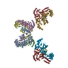
| ||||||||||||||||
|---|---|---|---|---|---|---|---|---|---|---|---|---|---|---|---|---|---|
| 1 | 
| ||||||||||||||||
| 2 | 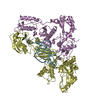
| ||||||||||||||||
| 3 | 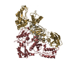
| ||||||||||||||||
| 4 | 
| ||||||||||||||||
| Unit cell |
| ||||||||||||||||
| Noncrystallographic symmetry (NCS) | NCS oper:
| ||||||||||||||||
| Details | THE HIV-1 REVERSE TRANSCRIPTASE IS A HETERODIMER CONSISTING OF TWO SUBUNITS: P66 (CHAIN A) AND P51 (DESIGNATED CHAIN B) IN THIS CRYSTAL FORM THERE ARE FOUR REVERSE TRANSCRIPTASE MOLECULES PLUS FOUR RNA MOLECULES PER ASYMMETRIC UNIT. THE COORDINATES FOR ONE COMPLEX ARE CONTAINED IN THIS ENTRY. THE NON-CRYSTALLOGRAPHIC SYMMETRY TRANSFORMATIONS GIVEN BELOW ARE BASED ON THE BEST SUPERPOSITION OF THIS MOLECULE ON THE OTHER THREE HETERODIMER COMPLEXES. THE TRANSFORMATIONS MATRICES IN THE MTRIX RECORDS BELOW DESCRIBE NON-CRYSTALLOGRAPHIC SYMMETRY AMONG THE FOUR HETERODIMERS OF REVERSE TRANSCRIPTASE FOUND IN THE ASYMMETRIC UNIT. APPLYING THE APPROPRIATE MTRIX TRANSFORMATION TO THE RESIDUES LISTED FIRST WILL YIELD COORDINATES FOR THE OTHER THREE MOLECULES PRESENT IN THE ASYMMETRIC UNIT. |
-Components
| #1: RNA chain | Mass: 9638.777 Da / Num. of mol.: 4 / Source method: obtained synthetically #2: Protein | Mass: 63834.125 Da / Num. of mol.: 4 Source method: isolated from a genetically manipulated source Details: THE HIV-1 REVERSE TRANSCRIPTASE IS A HETERODIMER CONSISTING OF TWO SUBUNITS: P66 (CHAIN A) AND P51 (DESIGNATED CHAIN B) IN THIS CRYSTAL FORM THERE ARE FOUR REVERSE TRANSCRIPTASE MOLECULES ...Details: THE HIV-1 REVERSE TRANSCRIPTASE IS A HETERODIMER CONSISTING OF TWO SUBUNITS: P66 (CHAIN A) AND P51 (DESIGNATED CHAIN B) IN THIS CRYSTAL FORM THERE ARE FOUR REVERSE TRANSCRIPTASE MOLECULES PLUS FOUR RNA MOLECULES PER ASYMMETRIC UNIT. THE COORDINATES FOR ONE COMPLEX ARE CONTAINED IN THIS ENTRY. THE NON-CRYSTALLOGRAPHIC SYMMETRY TRANSFORMATIONS GIVEN BELOW ARE BASED ON THE BEST SUPERPOSITION OF THIS MOLECULE ON THE OTHER THREE HETERODIMER COMPLEXES. Source: (gene. exp.) Human immunodeficiency virus 1 / Genus: Lentivirus / Variant: BH10 ISOLATE / Production host:  #3: Protein | Mass: 49532.961 Da / Num. of mol.: 4 Source method: isolated from a genetically manipulated source Details: THE HIV-1 REVERSE TRANSCRIPTASE IS A HETERODIMER CONSISTING OF TWO SUBUNITS: P66 (CHAIN A) AND P51 (DESIGNATED CHAIN B) IN THIS CRYSTAL FORM THERE ARE FOUR REVERSE TRANSCRIPTASE MOLECULES ...Details: THE HIV-1 REVERSE TRANSCRIPTASE IS A HETERODIMER CONSISTING OF TWO SUBUNITS: P66 (CHAIN A) AND P51 (DESIGNATED CHAIN B) IN THIS CRYSTAL FORM THERE ARE FOUR REVERSE TRANSCRIPTASE MOLECULES PLUS FOUR RNA MOLECULES PER ASYMMETRIC UNIT. THE COORDINATES FOR ONE COMPLEX ARE CONTAINED IN THIS ENTRY. THE NON-CRYSTALLOGRAPHIC SYMMETRY TRANSFORMATIONS GIVEN BELOW ARE BASED ON THE BEST SUPERPOSITION OF THIS MOLECULE ON THE OTHER THREE HETERODIMER COMPLEXES. Source: (gene. exp.) Human immunodeficiency virus 1 / Genus: Lentivirus / Variant: BH10 ISOLATE / Production host: |
|---|
-Experimental details
-Experiment
| Experiment | Method: X-RAY DIFFRACTION / Number of used crystals: 1 |
|---|
- Sample preparation
| Crystal | Density Matthews: 3.8 Å3/Da / Density % sol: 72 % | ||||||||||||||||||||||||||||||||||||||||||||||||||
|---|---|---|---|---|---|---|---|---|---|---|---|---|---|---|---|---|---|---|---|---|---|---|---|---|---|---|---|---|---|---|---|---|---|---|---|---|---|---|---|---|---|---|---|---|---|---|---|---|---|---|---|
| Crystal grow | Temperature: 277 K / Method: vapor diffusion, hanging drop / pH: 7 Details: pH 7.0, VAPOR DIFFUSION, HANGING DROP, temperature 277K | ||||||||||||||||||||||||||||||||||||||||||||||||||
| Components of the solutions |
| ||||||||||||||||||||||||||||||||||||||||||||||||||
| Crystal grow | *PLUS Temperature: 4 ℃ | ||||||||||||||||||||||||||||||||||||||||||||||||||
| Components of the solutions | *PLUS
|
-Data collection
| Diffraction | Mean temperature: 100 K |
|---|---|
| Diffraction source | Source: SYNCHROTRON / Site: CHESS  / Beamline: A1 / Wavelength: 0.92 / Beamline: A1 / Wavelength: 0.92 |
| Detector | Type: CUSTOM-MADE / Detector: CCD / Date: Mar 15, 1995 / Details: MIRROR |
| Radiation | Monochromator: SI(111) / Protocol: SINGLE WAVELENGTH / Monochromatic (M) / Laue (L): M / Scattering type: x-ray |
| Radiation wavelength | Wavelength: 0.92 Å / Relative weight: 1 |
| Reflection | Resolution: 4.75→30 Å / Num. obs: 32343 / % possible obs: 81 % / Observed criterion σ(I): 1.1 / Redundancy: 1.9 % / Rsym value: 0.064 |
| Reflection | *PLUS Rmerge(I) obs: 0.064 |
| Reflection shell | *PLUS % possible obs: 56.9 % / Rmerge(I) obs: 0.263 |
- Processing
| Software |
| ||||||||||||||||||||||||||||||||||||||||||||||||||||||||||||
|---|---|---|---|---|---|---|---|---|---|---|---|---|---|---|---|---|---|---|---|---|---|---|---|---|---|---|---|---|---|---|---|---|---|---|---|---|---|---|---|---|---|---|---|---|---|---|---|---|---|---|---|---|---|---|---|---|---|---|---|---|---|
| Refinement | Method to determine structure: MOLECULAR REPLACEMENT Starting model: 3HVT AND 1HMV Resolution: 4.75→30 Å / Cross valid method: FREE R / σ(F): 1.1 Details: RIGID BODY REFINEMENT ONLY DUE TO LIMITED RESOLUTION. THE FOLLOWING RESIDUES ARE NOT VISIBLE IN THE ELECTRON DENSITY MAPS, THEREFORE THEY ARE NOT INCLUDED IN THE COORDINATE LIST. CHAIN B: 212 - 234, 428 - 440.
| ||||||||||||||||||||||||||||||||||||||||||||||||||||||||||||
| Displacement parameters | Biso mean: 55 Å2 | ||||||||||||||||||||||||||||||||||||||||||||||||||||||||||||
| Refinement step | Cycle: LAST / Resolution: 4.75→30 Å
| ||||||||||||||||||||||||||||||||||||||||||||||||||||||||||||
| Refine LS restraints |
| ||||||||||||||||||||||||||||||||||||||||||||||||||||||||||||
| Software | *PLUS Name: X-PLOR / Version: 3.853 / Classification: refinement | ||||||||||||||||||||||||||||||||||||||||||||||||||||||||||||
| Refinement | *PLUS | ||||||||||||||||||||||||||||||||||||||||||||||||||||||||||||
| Solvent computation | *PLUS | ||||||||||||||||||||||||||||||||||||||||||||||||||||||||||||
| Displacement parameters | *PLUS |
