 Movie
Movie Controller

[English] 日本語
 Yorodumi
- PDB-1qmc: C-terminal DNA-binding domain of HIV-1 integrase, NMR, 42 structures -
+ Open data

- Basic information
| Entry | Database: PDB / ID: 1qmc | ||||||
|---|---|---|---|---|---|---|---|
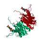
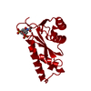
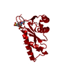
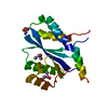
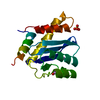
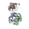
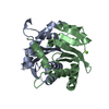
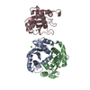
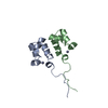
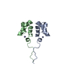
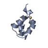
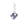
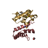
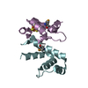
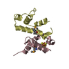
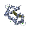
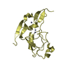
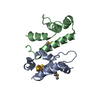
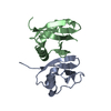
| Title | C-terminal DNA-binding domain of HIV-1 integrase, NMR, 42 structures | ||||||
 Components Components | HIV-1 INTEGRASE | ||||||
 Keywords Keywords | TRANSFERASE / INTEGRASE / DNA-BINDING PROTEIN / SRC HOMOLOGY 3 (SH3)-LIKE FOLD / AIDS / POLYPROTEIN | ||||||
| Function / homology |  Function and homology information Function and homology informationHIV-1 retropepsin / symbiont-mediated activation of host apoptosis / retroviral ribonuclease H / exoribonuclease H / exoribonuclease H activity / DNA integration / viral genome integration into host DNA / establishment of integrated proviral latency / RNA-directed DNA polymerase / RNA stem-loop binding ...HIV-1 retropepsin / symbiont-mediated activation of host apoptosis / retroviral ribonuclease H / exoribonuclease H / exoribonuclease H activity / DNA integration / viral genome integration into host DNA / establishment of integrated proviral latency / RNA-directed DNA polymerase / RNA stem-loop binding / viral penetration into host nucleus / host multivesicular body / RNA-directed DNA polymerase activity / RNA-DNA hybrid ribonuclease activity / Transferases; Transferring phosphorus-containing groups; Nucleotidyltransferases / host cell / viral nucleocapsid / DNA recombination / DNA-directed DNA polymerase / aspartic-type endopeptidase activity / Hydrolases; Acting on ester bonds / DNA-directed DNA polymerase activity / symbiont-mediated suppression of host gene expression / viral translational frameshifting / symbiont entry into host cell / lipid binding / host cell nucleus / host cell plasma membrane / virion membrane / structural molecule activity / proteolysis / DNA binding / zinc ion binding Similarity search - Function | ||||||
| Biological species |  HUMAN IMMUNODEFICIENCY VIRUS TYPE 1 BH10 HUMAN IMMUNODEFICIENCY VIRUS TYPE 1 BH10 | ||||||
| Method | SOLUTION NMR / simulated annealing | ||||||
 Authors Authors | Eijkelenboom, A.P.A.M. / Sprangers, R. / Hard, K. / Puras Lutzke, R.A. / Plasterk, R.H.A. / Boelens, R. / Kaptein, R. | ||||||
 Citation Citation | Journal: Proteins: Struct.,Funct., Genet. / Year: 1999 Title: Refined Solution Structure of the C-Terminal DNA-Binding Domain of Human Immunovirus-1 Integrase. Authors: Eijkelenboom, A.P.A.M. / Sprangers, R. / Hard, K. / Puras Lutzke, R.A. / Plasterk, R.H.A. / Boelens, R. / Kaptein, R. #1: Journal: Nat.Struct.Biol. / Year: 1995 Title: The DNA-Binding Domain of HIV-1 Integrase Has an SH3-Like Fold Authors: Eijkelenboom, A.P.A.M. / Puras Lutzke, R.A. / Boelens, R. / Plasterk, R.H.A. / Kaptein, R. / Hard, K. | ||||||
| History |
| ||||||
| Remark 700 | SHEET THE FIVE-STRANDED SHEET STRUCTURE OF THIS MOLECULE CONTAINS TWO BIFURCATED SHEETS IN THIS ... SHEET THE FIVE-STRANDED SHEET STRUCTURE OF THIS MOLECULE CONTAINS TWO BIFURCATED SHEETS IN THIS STRUCTURE. EACH IS REPRESENTED BY SHEETS WHICH HAVE ONE OR MORE IDENTICAL STRANDS. SHEETS AB AND AC (CHAIN A) REPRESENT THE BIFURCATED CONTRIBUTIONS TO THE MAIN 5-STRANDED SHEET STRUCTURE. |
- Structure visualization
| Structure viewer | Molecule: MolmilJmol/JSmol |
|---|
- Downloads & links
-Download
| PDBx/mmCIF format | 1qmc.cif.gz | 1.4 MB | Display | PDBx/mmCIF format |
|---|---|---|---|---|
| PDB format | pdb1qmc.ent.gz | 1.2 MB | Display | PDB format |
| PDBx/mmJSON format | 1qmc.json.gz | Tree view | PDBx/mmJSON format | |
| Others |  Other downloads Other downloads |
-Validation report
| Arichive directory | https://data.pdbj.org/pub/pdb/validation_reports/qm/1qmcftp://data.pdbj.org/pub/pdb/validation_reports/qm/1qmc | HTTPS FTP |
|---|
-Related structure data
| Related structure data | |
|---|---|
| Similar structure data |


-Links
 PDBj
PDBj


- Assembly
| Deposited unit | 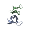
| |||||||||
|---|---|---|---|---|---|---|---|---|---|---|
| 1 |
| |||||||||
| NMR ensembles |
|
-Components
| #1: Protein | Mass: 6152.228 Da / Num. of mol.: 2 / Fragment: C-TERMINAL DNA-BINDING DOMAIN Source method: isolated from a genetically manipulated source Source: (gene. exp.) HUMAN IMMUNODEFICIENCY VIRUS TYPE 1 BH10Production host:  |
|---|
-Experimental details
-Experiment
| Experiment | Method: SOLUTION NMR |
|---|---|
| NMR details | Text: MODEL 1 IS CLOSEST TO THE AVERAGE FOR RESIDUES 220-270. RESIDUE 219 IS DISORDERED. |
- Sample preparation
| Sample conditions | pH: 4.7 / Temperature: 298 K |
|---|---|
| Crystal grow | *PLUS Method: other / Details: NMR |
-NMR measurement
| NMR spectrometer |
|
|---|
- Processing
| NMR software |
| ||||||||||||
|---|---|---|---|---|---|---|---|---|---|---|---|---|---|
| Refinement | Method: simulated annealing / Software ordinal: 1 Details: REFINEMENT DETAILS CAN BE FOUND IN THE JRNL CITATION ABOVE. | ||||||||||||
| NMR ensemble | Conformer selection criteria: LOW OVERALL ENERGY / Conformers calculated total number: 50 / Conformers submitted total number: 42 |
 X-PLOR