 Movie
Movie Controller

[English] 日本語
 Yorodumi
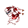
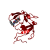
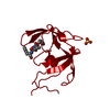
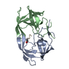
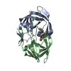
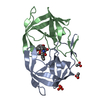
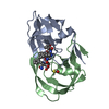
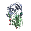
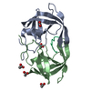
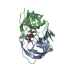
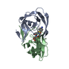
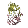
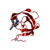
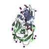
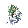
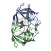
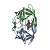
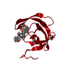
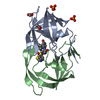
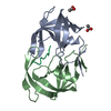
- PDB-3tlh: STRUCTURAL STUDIES OF HIV AND FIV PROTEASES COMPLEXED WITHAN EFFI... -
+ Open data

- Basic information
| Entry | Database: PDB / ID: 3tlh | ||||||
|---|---|---|---|---|---|---|---|
| Title | STRUCTURAL STUDIES OF HIV AND FIV PROTEASES COMPLEXED WITHAN EFFICIENT INHIBITOR OF FIV PR | ||||||
 Components Components | PROTEIN (PROTEASE) | ||||||
 Keywords Keywords | HYDROLASE/HYDROLASE INHIBITOR / HIV PROTEASE / HYDROLASE-HYDROLASE INHIBITOR COMPLEX | ||||||
| Function / homology |  Function and homology information Function and homology informationHIV-1 retropepsin / symbiont-mediated activation of host apoptosis / retroviral ribonuclease H / exoribonuclease H / exoribonuclease H activity / DNA integration / viral genome integration into host DNA / establishment of integrated proviral latency / RNA-directed DNA polymerase / RNA stem-loop binding ...HIV-1 retropepsin / symbiont-mediated activation of host apoptosis / retroviral ribonuclease H / exoribonuclease H / exoribonuclease H activity / DNA integration / viral genome integration into host DNA / establishment of integrated proviral latency / RNA-directed DNA polymerase / RNA stem-loop binding / viral penetration into host nucleus / host multivesicular body / RNA-directed DNA polymerase activity / RNA-DNA hybrid ribonuclease activity / Transferases; Transferring phosphorus-containing groups; Nucleotidyltransferases / host cell / viral nucleocapsid / DNA recombination / DNA-directed DNA polymerase / aspartic-type endopeptidase activity / Hydrolases; Acting on ester bonds / DNA-directed DNA polymerase activity / symbiont-mediated suppression of host gene expression / viral translational frameshifting / symbiont entry into host cell / lipid binding / host cell nucleus / host cell plasma membrane / virion membrane / structural molecule activity / proteolysis / DNA binding / zinc ion binding Similarity search - Function | ||||||
| Biological species |   Human immunodeficiency virus type 1 Human immunodeficiency virus type 1 | ||||||
| Method |  X-RAY DIFFRACTION / OTHER / Resolution: 2 Å X-RAY DIFFRACTION / OTHER / Resolution: 2 Å | ||||||
 Authors Authors | Li, M. / Lee, T. / Morris, G. / Laco, G. / Wong, C. / Olson, A. / Elder, J. / Wlodawer, A. / Gustchina, A. | ||||||
 Citation Citation | Journal: Proteins / Year: 2000 Title: Structural studies of FIV and HIV-1 proteases complexed with an efficient inhibitor of FIV protease Authors: Li, M. / Morris, G.M. / Lee, T. / Laco, G.S. / Wong, C.H. / Olson, A.J. / Elder, J.H. / Wlodawer, A. / Gustchina, A. #1: Journal: Nat.Struct.Biol. / Year: 1995Title: Structural Studies of HIV and Fiv Proteases Complexed with an Efficient Inhibitor of Fiv Pr Authors: Wlodawer, A. / Gustchina, A. / Reshetnikova, L. / Lubkowski, J. / Zdanov, A. / Hui, K. / Angleton, E. / Farmerie, W. / Goodenow, M. / Bhatt, D. / Zhang, L. / Dunn, B. | ||||||
| History |
|
- Structure visualization
| Structure viewer | Molecule: MolmilJmol/JSmol |
|---|
- Downloads & links
-Download
| PDBx/mmCIF format | 3tlh.cif.gz | 35.1 KB | Display | PDBx/mmCIF format |
|---|---|---|---|---|
| PDB format | pdb3tlh.ent.gz | 22.4 KB | Display | PDB format |
| PDBx/mmJSON format | 3tlh.json.gz | Tree view | PDBx/mmJSON format | |
| Others |  Other downloads Other downloads |
-Validation report
| Arichive directory | https://data.pdbj.org/pub/pdb/validation_reports/tl/3tlhftp://data.pdbj.org/pub/pdb/validation_reports/tl/3tlh | HTTPS FTP |
|---|
-Related structure data


-Links
 PDBj
PDBj


- Assembly
| Deposited unit | 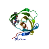
| |||||||||
|---|---|---|---|---|---|---|---|---|---|---|
| 1 | 
| |||||||||
| Unit cell |
| |||||||||
| Components on special symmetry positions |
|
-Components
| #1: Protein | Mass: 10804.808 Da / Num. of mol.: 1 Source method: isolated from a genetically manipulated source Details: COMPLEXED WITH TL-3-093 Source: (gene. exp.) Human immunodeficiency virus type 1 (CLONE 12)Genus: Lentivirus / Species: Human immunodeficiency virus 1 / Production host:  |
|---|---|
| #2: Chemical | ChemComp-3TL / 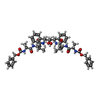  Type: peptide-like, Peptide-like / Class: Inhibitor / Mass: 909.077 Da / Num. of mol.: 1 / Source method: obtained synthetically / Formula: C50H64N6O10 Type: peptide-like, Peptide-like / Class: Inhibitor / Mass: 909.077 Da / Num. of mol.: 1 / Source method: obtained synthetically / Formula: C50H64N6O10References: N-[(benzyloxy)carbonyl]-L-alanyl-N-[(1R)-1-benzyl-2-oxoethyl]-L-valinamide |
| #3: Water | ChemComp-HOH /  Mass: 18.015 Da / Num. of mol.: 33 / Source method: isolated from a natural source / Formula: H2O Mass: 18.015 Da / Num. of mol.: 33 / Source method: isolated from a natural source / Formula: H2O |
| Nonpolymer details | THE INHIBITOR IS A C2 SYMMETRIC HIV PROTEASE |
-Experimental details
-Experiment
| Experiment | Method: X-RAY DIFFRACTION / Number of used crystals: 1 |
|---|
- Sample preparation
| Crystal | Density Matthews: 2.2 Å3/Da / Density % sol: 44.4 % | ||||||||||||||||||||||||||||||
|---|---|---|---|---|---|---|---|---|---|---|---|---|---|---|---|---|---|---|---|---|---|---|---|---|---|---|---|---|---|---|---|
| Crystal grow | pH: 5.4 Details: SAMPLE: 3.0MG/ML HIVPR IN 25MM NAACETATE WITH 1MM EDTA AND 10MM DTT AT PH 5.5. WELL SOLUTION: 1.3M AMMONIUM SULFATE IN SODIUM ACETATE BUFFER AT PH 5.3 MIXING SAMPLE AND WELL SOLLUTION 1:1, pH 5.4 PH range: 5.3-5.5 | ||||||||||||||||||||||||||||||
| Crystal grow | *PLUS pH: 6.2 / Method: vapor diffusion, hanging drop | ||||||||||||||||||||||||||||||
| Components of the solutions | *PLUS
|
-Data collection
| Diffraction | Mean temperature: 293 K |
|---|---|
| Diffraction source | Source: ROTATING ANODE / Type: RIGAKU RU200 / Wavelength: 1.5418 |
| Detector | Type: MAR scanner 345 mm plate / Detector: IMAGE PLATE / Date: Feb 1, 1998 / Details: MIRRORS |
| Radiation | Protocol: SINGLE WAVELENGTH / Monochromatic (M) / Laue (L): M / Scattering type: x-ray |
| Radiation wavelength | Wavelength: 1.5418 Å / Relative weight: 1 |
| Reflection | Resolution: 1.9→20 Å / Num. obs: 8126 / % possible obs: 98.4 % / Observed criterion σ(I): 0 / Redundancy: 6.1 % / Rmerge(I) obs: 0.106 / Net I/σ(I): 10.5 |
| Reflection shell | Resolution: 1.9→1.93 Å / Mean I/σ(I) obs: 2.6 / Rsym value: 0.419 / % possible all: 96.2 |
| Reflection shell | *PLUS % possible obs: 96.2 % / Rmerge(I) obs: 0.419 |
- Processing
| Software |
| |||||||||||||||||||||||||||||||||
|---|---|---|---|---|---|---|---|---|---|---|---|---|---|---|---|---|---|---|---|---|---|---|---|---|---|---|---|---|---|---|---|---|---|---|
| Refinement | Method to determine structure: OTHER / Resolution: 2→10 Å / Num. parameters: 3323 / Num. restraintsaints: 3233 / Cross valid method: THROUGHOUT / σ(F): 0 / Stereochemistry target values: ENGH AND HUBER Details: ANISOTROPIC SCALING APPLIED BY METHOD PF PARKIN, MOEZZI & HOPE, J.APPL.CRYST.28(1995)53-56
| |||||||||||||||||||||||||||||||||
| Solvent computation | Solvent model: MOEWS & KRETSINGER | |||||||||||||||||||||||||||||||||
| Refine analyze | Num. disordered residues: 1 / Occupancy sum hydrogen: 0 / Occupancy sum non hydrogen: 819.5 | |||||||||||||||||||||||||||||||||
| Refinement step | Cycle: LAST / Resolution: 2→10 Å
| |||||||||||||||||||||||||||||||||
| Refine LS restraints |
| |||||||||||||||||||||||||||||||||
| Software | *PLUS Name: SHELXL-97 / Classification: refinement | |||||||||||||||||||||||||||||||||
| Refinement | *PLUS Rfactor obs: 0.195 / Rfactor Rfree: 0.281 / Rfactor Rwork: 0.186 | |||||||||||||||||||||||||||||||||
| Solvent computation | *PLUS | |||||||||||||||||||||||||||||||||
| Displacement parameters | *PLUS Biso mean: 34.53 Å2 | |||||||||||||||||||||||||||||||||
| Refine LS restraints | *PLUS
|
