 Movie
Movie Controller

[English] 日本語
 Yorodumi
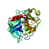
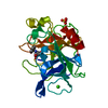
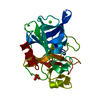
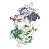
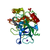
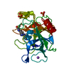
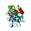
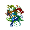
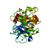
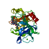
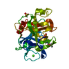
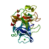
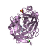
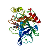
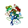
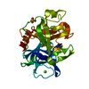
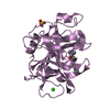
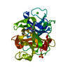
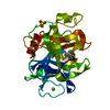
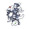
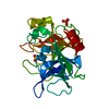
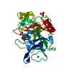
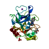
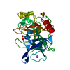
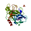
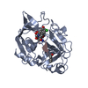
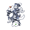
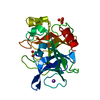
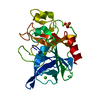
- PDB-1h9l: PORCINE PANCREATIC ELASTASE COMPLEXED WITH ACETYL-VAL-GLU-PRO-ILE-COOH -
+ Open data

- Basic information
| Entry | Database: PDB / ID: 1h9l | ||||||
|---|---|---|---|---|---|---|---|
| Title | PORCINE PANCREATIC ELASTASE COMPLEXED WITH ACETYL-VAL-GLU-PRO-ILE-COOH | ||||||
 Components Components |
| ||||||
 Keywords Keywords | HYDROLASE/HYDROLASE INHIBITOR / SERINE PROTEINASE / HYDROLASE / HYDROLASE-HYDROLASE INHIBITOR COMPLEX | ||||||
| Function / homology |  Function and homology information Function and homology informationpancreatic elastase / serine-type endopeptidase activity / proteolysis / : / metal ion binding Similarity search - Function | ||||||
| Biological species |  synthetic construct (others) | ||||||
| Method |  X-RAY DIFFRACTION / SYNCHROTRON / MOLECULAR REPLACEMENT / Resolution: 1.67 Å X-RAY DIFFRACTION / SYNCHROTRON / MOLECULAR REPLACEMENT / Resolution: 1.67 Å | ||||||
 Authors Authors | Wright, P.A. / Wilmouth, R.C. / Clifton, I.J. / Schofield, C.J. | ||||||
 Citation Citation | Journal: Eur.J.Biochem. / Year: 2001 Title: Kinetic and Crystallographic Analysis of Complexes Formed between Elastase and Peptides from Beta-Casein Authors: Wright, P.A. / Wilmouth, R.C. / Clifton, I.J. / Schofield, C.J. #1: Journal: Nat.Struct.Biol. / Year: 1997Title: Structure of a Specific Acyl-Enzyme Complex Formed between Beta-Casomorphin-7 and Porcine Pancreatic Elastase Authors: Wilmouth, R.C. / Clifton, I.J. / Robinson, C.V. / Roach, P.L. / Aplin, R.T. / Westwood, N.J. / Hajdu, J. / Schofield, C.J. #2: Journal: Acta Crystallogr.,Sect.B / Year: 1988Title: Structure of Native Porcine Pancreatic Elastase at 1.65A Resolution Authors: Meyer, E. / Cole, G. / Radhakrishnan, R. / Epp, O. #3: Journal: Biochem.J. / Year: 2000Title: Ph-Jump Crystallographic Analyses of Gamma-Lactam-Porcine Pancreatic Elastase Complexes Authors: Wright, P.A. / Wilmouth, R.C. / Clifton, I.J. / Schofield, C.J. | ||||||
| History |
| ||||||
| Remark 700 | SHEET DETERMINATION METHOD: DSSP THE SHEETS PRESENTED AS "B" IN EACH CHAIN ON SHEET RECORDS BELOW ... SHEET DETERMINATION METHOD: DSSP THE SHEETS PRESENTED AS "B" IN EACH CHAIN ON SHEET RECORDS BELOW IS ACTUALLY AN 6-STRANDED BARREL THIS IS REPRESENTED BY A 7-STRANDED SHEET IN WHICH THE FIRST AND LAST STRANDS ARE IDENTICAL. |
- Structure visualization
| Structure viewer | Molecule: MolmilJmol/JSmol |
|---|
- Downloads & links
-Download
| PDBx/mmCIF format | 1h9l.cif.gz | 67.4 KB | Display | PDBx/mmCIF format |
|---|---|---|---|---|
| PDB format | pdb1h9l.ent.gz | 48.4 KB | Display | PDB format |
| PDBx/mmJSON format | 1h9l.json.gz | Tree view | PDBx/mmJSON format | |
| Others |  Other downloads Other downloads |
-Validation report
| Arichive directory | https://data.pdbj.org/pub/pdb/validation_reports/h9/1h9lftp://data.pdbj.org/pub/pdb/validation_reports/h9/1h9l | HTTPS FTP |
|---|
-Related structure data
| Related structure data | 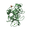 1qixS S: Starting model for refinement |
|---|---|
| Similar structure data |


-Links
 PDBj
PDBj
- Assembly
| Deposited unit | 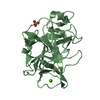
| ||||||||
|---|---|---|---|---|---|---|---|---|---|
| 1 |
| ||||||||
| Unit cell |
|
-Components
| #1: Protein/peptide | Mass: 482.570 Da / Num. of mol.: 1 / Source method: obtained synthetically / Source: (synth.) synthetic construct (others) |
|---|---|
| #2: Protein | Mass: 25928.031 Da / Num. of mol.: 1 / Source method: isolated from a natural source / Source: (natural) |
| #3: Chemical | ChemComp-CA /   Mass: 40.078 Da / Num. of mol.: 1 / Source method: obtained synthetically / Formula: Ca Mass: 40.078 Da / Num. of mol.: 1 / Source method: obtained synthetically / Formula: Ca |
| #4: Chemical | ChemComp-SO4 /   Mass: 96.063 Da / Num. of mol.: 1 / Source method: obtained synthetically / Formula: SO4 Mass: 96.063 Da / Num. of mol.: 1 / Source method: obtained synthetically / Formula: SO4 |
| #5: Water | ChemComp-HOH /  Mass: 18.015 Da / Num. of mol.: 233 / Source method: isolated from a natural source / Formula: H2O Mass: 18.015 Da / Num. of mol.: 233 / Source method: isolated from a natural source / Formula: H2O |
| Compound details | THERE IS AN ESTER BOND BETWEEN THE CARBOXYL TERMINUS OF THE ACETYL-VAL-GLU-PRO-ILE PEPTIDE AND THE ...THERE IS AN ESTER BOND BETWEEN THE CARBOXYL TERMINUS OF THE ACETYL-VAL-GLU-PRO-ILE PEPTIDE AND THE HYDROXYL SIDE CHAIN OF SER-195 |
| Has protein modification | Y |
-Experimental details
-Experiment
| Experiment | Method: X-RAY DIFFRACTION / Number of used crystals: 1 |
|---|
- Sample preparation
| Crystal | Density Matthews: 1.83 Å3/Da / Density % sol: 32.15 % | ||||||||||||||||||||||||||||
|---|---|---|---|---|---|---|---|---|---|---|---|---|---|---|---|---|---|---|---|---|---|---|---|---|---|---|---|---|---|
| Crystal grow | pH: 5 Details: CRYSTALS PREPARED FROM 25MM SODIUM SULPHATE, 25MM SODIUM ACETATE (PH 5.0), 25 MG/ML PPE AND THEN SOAKED WITH ACETYL-VAL-GLU-PRO-ILE (17 MG/ML) DISSOLVED IN SODIUM ACETATE (PH 5.0, 50MM) AND ...Details: CRYSTALS PREPARED FROM 25MM SODIUM SULPHATE, 25MM SODIUM ACETATE (PH 5.0), 25 MG/ML PPE AND THEN SOAKED WITH ACETYL-VAL-GLU-PRO-ILE (17 MG/ML) DISSOLVED IN SODIUM ACETATE (PH 5.0, 50MM) AND SODIUM SULPHATE (25 MM) | ||||||||||||||||||||||||||||
| Crystal grow | *PLUS Temperature: 17 ℃ / Method: vapor diffusion, hanging drop | ||||||||||||||||||||||||||||
| Components of the solutions | *PLUS
|
-Data collection
| Diffraction | Mean temperature: 100 K |
|---|---|
| Diffraction source | Source: SYNCHROTRON / Site: SRS  / Beamline: PX9.6 / Wavelength: 0.87 / Beamline: PX9.6 / Wavelength: 0.87 |
| Detector | Type: MARRESEARCH / Detector: IMAGE PLATE / Date: Jun 15, 1998 |
| Radiation | Protocol: SINGLE WAVELENGTH / Monochromatic (M) / Laue (L): M / Scattering type: x-ray |
| Radiation wavelength | Wavelength: 0.87 Å / Relative weight: 1 |
| Reflection | Resolution: 1.67→22.9 Å / Num. obs: 24944 / % possible obs: 95.2 % / Observed criterion σ(I): 0 / Redundancy: 6.3 % / Biso Wilson estimate: 13.8 Å2 / Rmerge(I) obs: 0.072 / Net I/σ(I): 15 |
| Reflection shell | Resolution: 1.67→1.7 Å / Rmerge(I) obs: 0.25 / Mean I/σ(I) obs: 10 / % possible all: 96.7 |
| Reflection | *PLUS Num. measured all: 156137 |
| Reflection shell | *PLUS % possible obs: 96.7 % |
- Processing
| Software |
| ||||||||||||||||||||||||||||||||||||||||||||||||||||||||||||||||||||||||||||||||||||
|---|---|---|---|---|---|---|---|---|---|---|---|---|---|---|---|---|---|---|---|---|---|---|---|---|---|---|---|---|---|---|---|---|---|---|---|---|---|---|---|---|---|---|---|---|---|---|---|---|---|---|---|---|---|---|---|---|---|---|---|---|---|---|---|---|---|---|---|---|---|---|---|---|---|---|---|---|---|---|---|---|---|---|---|---|---|
| Refinement | Method to determine structure: MOLECULAR REPLACEMENT Starting model: PDB ENTRY 1QIX Resolution: 1.67→22.9 Å / SU B: 1.841 / SU ML: 0.064 / Cross valid method: THROUGHOUT / σ(F): 0 / ESU R: 0.1 / ESU R Free: 0.102 Details: THE STRUCTURE WAS REFINED WITHOUT ANGLE OR PLANARITY RESTRAINTS ON THE ESTER BOND BETWEEN SER- 195 AND THE PEPTIDE ISOLEUCINE.THE N-TERMINAL ACETYL GROUP OF THE PEPTIDE WAS DISORDERED AND ...Details: THE STRUCTURE WAS REFINED WITHOUT ANGLE OR PLANARITY RESTRAINTS ON THE ESTER BOND BETWEEN SER- 195 AND THE PEPTIDE ISOLEUCINE.THE N-TERMINAL ACETYL GROUP OF THE PEPTIDE WAS DISORDERED AND HENCE IS MISSING FROM THE STRUCTURE
| ||||||||||||||||||||||||||||||||||||||||||||||||||||||||||||||||||||||||||||||||||||
| Displacement parameters | Biso mean: 15.5 Å2
| ||||||||||||||||||||||||||||||||||||||||||||||||||||||||||||||||||||||||||||||||||||
| Refinement step | Cycle: LAST / Resolution: 1.67→22.9 Å
| ||||||||||||||||||||||||||||||||||||||||||||||||||||||||||||||||||||||||||||||||||||
| Refine LS restraints |
| ||||||||||||||||||||||||||||||||||||||||||||||||||||||||||||||||||||||||||||||||||||
| Refine LS restraints | *PLUS
|
