 Movie
Movie Controller

[English] 日本語
 Yorodumi
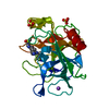
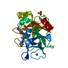
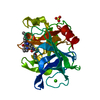
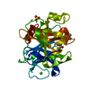
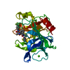
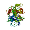
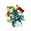
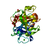
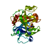
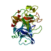
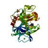
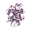
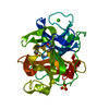
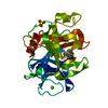
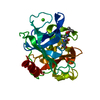
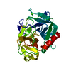
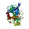
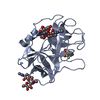
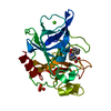
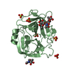
- PDB-1qnj: THE STRUCTURE OF NATIVE PORCINE PANCREATIC ELASTASE AT ATOMIC RES... -
+ Open data

- Basic information
| Entry | Database: PDB / ID: 1qnj | ||||||
|---|---|---|---|---|---|---|---|
| Title | THE STRUCTURE OF NATIVE PORCINE PANCREATIC ELASTASE AT ATOMIC RESOLUTION (1.1 A) | ||||||
 Components Components | ELASTASE | ||||||
 Keywords Keywords | HYDROLASE (SERINE PROTEASE) / HYDROLASE(SERINE PROTEASE) / ATOMIC RESOLUTION | ||||||
| Function / homology |  Function and homology information Function and homology informationpancreatic elastase / serine-type endopeptidase activity / proteolysis / : / metal ion binding Similarity search - Function | ||||||
| Biological species |  | ||||||
| Method |  X-RAY DIFFRACTION / SYNCHROTRON / MOLECULAR REPLACEMENT / Resolution: 1.1 Å X-RAY DIFFRACTION / SYNCHROTRON / MOLECULAR REPLACEMENT / Resolution: 1.1 Å | ||||||
 Authors Authors | Wurtele, M. / Hahn, M. / Hilpert, K. / Hohne, W. | ||||||
 Citation Citation | Journal: Acta Crystallogr.,Sect.D / Year: 2000 Title: Atomic Resolution Structure of Native Porcine Pancreatic Elastase at 1.1 A Authors: Wurtele, M. / Hahn, M. / Hilpert, K. / Hohne, W. #1: Journal: Biochemistry / Year: 1998Title: Inhibition of Elastase by N-Sulfonylaryl Beta-Lactams: Anatomy of a Stable Acyl-Enzyme Complex Authors: Wilmouth, R.C. / Westwood, N.J. / Anderson, K. / Brownlee, W. / Claridge, T.D.W. / Clifton, I.J. / Pritchard, G.J. / Aplin, R.T. / Schofield, C.J. #2: Journal: Acta Crystallogr.,Sect.B / Year: 1988Title: Structure of Native Porcine Pancreatic Elastase at 1.65 Angstroms Resolution Authors: Meyer, E. / Cole, G. / Radhakrishnan, R. / Epp, O. #3: Journal: J.Mol.Biol. / Year: 1978Title: The Atomic Structure of Crystalline Porcine Pancreatic Elastase at 2.5 Angstroms Resolution. Comparisons with the Structure of Alpha- Chymotrypsin Authors: Sawyer, L. / Shotton, D.M. / Campbell, J.W. / Wendell, P.L. / Muirhead, H. / Watson, H.C. / Diamond, R. / Ladner, R.C. #4: Journal: Nature / Year: 1970 Title: Three-Dimensional Fourier Synthesis of Tosyl-Elastase at 3.5 Angstroms Resolution Authors: Watson, H.C. / Shotton, D.M. / Cox, J.M. / Muirhead, H. | ||||||
| History |
|
- Structure visualization
| Structure viewer | Molecule: MolmilJmol/JSmol |
|---|
- Downloads & links
-Download
| PDBx/mmCIF format | 1qnj.cif.gz | 182.3 KB | Display | PDBx/mmCIF format |
|---|---|---|---|---|
| PDB format | pdb1qnj.ent.gz | 140.7 KB | Display | PDB format |
| PDBx/mmJSON format | 1qnj.json.gz | Tree view | PDBx/mmJSON format | |
| Others |  Other downloads Other downloads |
-Validation report
| Arichive directory | https://data.pdbj.org/pub/pdb/validation_reports/qn/1qnjftp://data.pdbj.org/pub/pdb/validation_reports/qn/1qnj | HTTPS FTP |
|---|
-Related structure data
| Related structure data |  1btuS S: Starting model for refinement |
|---|---|
| Similar structure data |


-Links
 PDBj
PDBj
- Assembly
| Deposited unit | 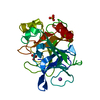
| ||||||||
|---|---|---|---|---|---|---|---|---|---|
| 1 |
| ||||||||
| Unit cell |
|
-Components
| #1: Protein | Mass: 25929.016 Da / Num. of mol.: 1 / Source method: isolated from a natural source / Details: PORCINE PANCREATIC ELASTASE / Source: (natural) | ||||||
|---|---|---|---|---|---|---|---|
| #2: Chemical | ChemComp-NA /   Mass: 22.990 Da / Num. of mol.: 1 / Source method: obtained synthetically / Formula: Na Mass: 22.990 Da / Num. of mol.: 1 / Source method: obtained synthetically / Formula: Na | ||||||
| #3: Chemical |   Mass: 96.063 Da / Num. of mol.: 2 / Source method: obtained synthetically / Formula: SO4 Mass: 96.063 Da / Num. of mol.: 2 / Source method: obtained synthetically / Formula: SO4#4: Water | ChemComp-HOH / |  Mass: 18.015 Da / Num. of mol.: 364 / Source method: isolated from a natural source / Formula: H2O Mass: 18.015 Da / Num. of mol.: 364 / Source method: isolated from a natural source / Formula: H2OCompound details | RESIDUE NUMBERING FOLLOWS THE NUMBERING OF BOVINE CHYMOTRYPSINOGEN A. THE RESIDUE ASN-77 OF ...RESIDUE NUMBERING FOLLOWS THE NUMBERING OF BOVINE CHYMOTRYPS | Has protein modification | Y | |
-Experimental details
-Experiment
| Experiment | Method: X-RAY DIFFRACTION / Number of used crystals: 1 |
|---|
- Sample preparation
| Crystal | Density Matthews: 2.06 Å3/Da / Density % sol: 32.6 % | ||||||||||||||||||
|---|---|---|---|---|---|---|---|---|---|---|---|---|---|---|---|---|---|---|---|
| Crystal grow | Temperature: 291 K / pH: 8 / Details: 0.25 M NA2SO4 18 DEGREES CELSIUS, pH 8.00 | ||||||||||||||||||
| Crystal grow | *PLUS Method: vapor diffusion, hanging drop / PH range low: 8 / PH range high: 7 | ||||||||||||||||||
| Components of the solutions | *PLUS
|
-Data collection
| Diffraction | Mean temperature: 100 K |
|---|---|
| Diffraction source | Source: SYNCHROTRON / Site: EMBL/DESY, HAMBURG  / Beamline: X11 / Wavelength: 0.9116 / Beamline: X11 / Wavelength: 0.9116 |
| Detector | Type: MARRESEARCH / Detector: IMAGE PLATE / Date: Mar 15, 1999 |
| Radiation | Protocol: SINGLE WAVELENGTH / Monochromatic (M) / Laue (L): M / Scattering type: x-ray |
| Radiation wavelength | Wavelength: 0.9116 Å / Relative weight: 1 |
| Reflection | Resolution: 1.1→45.6 Å / Num. obs: 81820 / % possible obs: 92.7 % / Redundancy: 13.1 % / Rmerge(I) obs: 0.045 / Net I/σ(I): 14.9 |
| Reflection shell | Resolution: 1.1→1.14 Å / Rmerge(I) obs: 0.35 / Mean I/σ(I) obs: 2.02 / % possible all: 60.4 |
| Reflection | *PLUS Num. measured all: 1071726 |
| Reflection shell | *PLUS % possible obs: 60.4 % |
- Processing
| Software |
| |||||||||||||||||||||||||||||||||
|---|---|---|---|---|---|---|---|---|---|---|---|---|---|---|---|---|---|---|---|---|---|---|---|---|---|---|---|---|---|---|---|---|---|---|
| Refinement | Method to determine structure: MOLECULAR REPLACEMENT Starting model: 1BTU Resolution: 1.1→45.6 Å / Num. parameters: 19897 / Num. restraintsaints: 24006 / Cross valid method: FREE R-VALUE / σ(F): 0 / Stereochemistry target values: ENGH AND HUBER Details: ANISOTROPIC SCALING APPLIED BY THE METHOD OF PARKIN, MOEZZI & HOPE, J.APPL.CRYST.28 (1995)53-56
| |||||||||||||||||||||||||||||||||
| Solvent computation | Solvent model: MOEWS & KRETSINGER | |||||||||||||||||||||||||||||||||
| Refine analyze | Num. disordered residues: 23 / Occupancy sum hydrogen: 1762 / Occupancy sum non hydrogen: 2177 | |||||||||||||||||||||||||||||||||
| Refinement step | Cycle: LAST / Resolution: 1.1→45.6 Å
| |||||||||||||||||||||||||||||||||
| Refine LS restraints |
| |||||||||||||||||||||||||||||||||
| Software | *PLUS Name: SHELXL-97 / Classification: refinement | |||||||||||||||||||||||||||||||||
| Refinement | *PLUS Rfactor Rwork: 0.1267 | |||||||||||||||||||||||||||||||||
| Solvent computation | *PLUS | |||||||||||||||||||||||||||||||||
| Displacement parameters | *PLUS | |||||||||||||||||||||||||||||||||
| Refine LS restraints | *PLUS Type: s_plane_restr / Dev ideal: 0.0289 |
