 Movie
Movie Controller

[English] 日本語
 Yorodumi
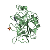
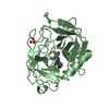
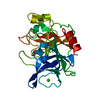
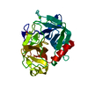
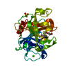
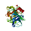
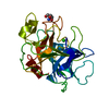
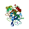
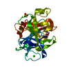
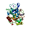
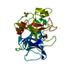
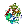
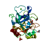
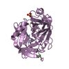
- PDB-2h1u: Porcine pancreatic elastase complexed with MetPheLeuGlu at pH 5.0 -
+ Open data

- Basic information
| Entry | Database: PDB / ID: 2h1u | ||||||
|---|---|---|---|---|---|---|---|
| Title | Porcine pancreatic elastase complexed with MetPheLeuGlu at pH 5.0 | ||||||
 Components Components |
| ||||||
 Keywords Keywords | HYDROLASE / SERINE PROTEINASE | ||||||
| Function / homology |  Function and homology information Function and homology informationpancreatic elastase / serine-type endopeptidase activity / proteolysis / : / metal ion binding Similarity search - Function | ||||||
| Biological species |  | ||||||
| Method |  X-RAY DIFFRACTION / MOLECULAR REPLACEMENT / Resolution: 1.6 Å X-RAY DIFFRACTION / MOLECULAR REPLACEMENT / Resolution: 1.6 Å | ||||||
 Authors Authors | Liu, B. / Schofield, C.J. / Wilmouth, R.C. | ||||||
 Citation Citation | Journal: J.Biol.Chem. / Year: 2006 Title: Structural analyses on intermediates in serine protease catalysis. Authors: Liu, B. / Schofield, C.J. / Wilmouth, R.C. #1: Journal: Bioorg.Med.Chem.Lett. / Year: 2000 Title: Mass spectrometry reveals elastase inhibitors from the reactive centre loop of alpha1-antitrypsin Authors: Wright, P.A. / Rostom, A.A. / Robinson, C.V. / Schofield, C.J. | ||||||
| History |
|
- Structure visualization
| Structure viewer | Molecule: MolmilJmol/JSmol |
|---|
- Downloads & links
-Download
| PDBx/mmCIF format | 2h1u.cif.gz | 62.6 KB | Display | PDBx/mmCIF format |
|---|---|---|---|---|
| PDB format | pdb2h1u.ent.gz | 44.3 KB | Display | PDB format |
| PDBx/mmJSON format | 2h1u.json.gz | Tree view | PDBx/mmJSON format | |
| Others |  Other downloads Other downloads |
-Validation report
| Arichive directory | https://data.pdbj.org/pub/pdb/validation_reports/h1/2h1uftp://data.pdbj.org/pub/pdb/validation_reports/h1/2h1u | HTTPS FTP |
|---|
-Related structure data
| Related structure data | 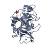 2bb4C 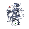 2bd2C 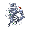 2bd3C 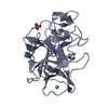 2bd4C  2bd5C 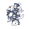 2bd7C 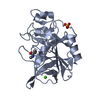 2bd8C 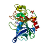 2bd9C 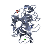 2bdaC 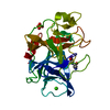 2bdbC 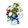 2bdcC 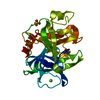 3estS C: citing same article ( S: Starting model for refinement |
|---|---|
| Similar structure data |


-Links
 PDBj
PDBj
- Assembly
| Deposited unit | 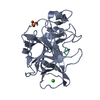
| ||||||||
|---|---|---|---|---|---|---|---|---|---|
| 1 |
| ||||||||
| Unit cell |
|
-Components
| #1: Protein/peptide | Mass: 538.656 Da / Num. of mol.: 1 / Source method: obtained synthetically / Details: synthetic peptide |
|---|---|
| #2: Protein | Mass: 25928.031 Da / Num. of mol.: 1 / Source method: isolated from a natural source / Source: (natural) |
| #3: Chemical | ChemComp-CA /   Mass: 40.078 Da / Num. of mol.: 1 / Source method: obtained synthetically / Formula: Ca Mass: 40.078 Da / Num. of mol.: 1 / Source method: obtained synthetically / Formula: Ca |
| #4: Chemical | ChemComp-SO4 /   Mass: 96.063 Da / Num. of mol.: 1 / Source method: obtained synthetically / Formula: SO4 Mass: 96.063 Da / Num. of mol.: 1 / Source method: obtained synthetically / Formula: SO4 |
| #5: Water | ChemComp-HOH /  Mass: 18.015 Da / Num. of mol.: 118 / Source method: isolated from a natural source / Formula: H2O Mass: 18.015 Da / Num. of mol.: 118 / Source method: isolated from a natural source / Formula: H2O |
| Has protein modification | Y |
-Experimental details
-Experiment
| Experiment | Method: X-RAY DIFFRACTION / Number of used crystals: 1 |
|---|
- Sample preparation
| Crystal | Density Matthews: 2.02 Å3/Da / Density % sol: 39.07 % |
|---|---|
| Crystal grow | Temperature: 294 K / Method: vapor diffusion, hanging drop / pH: 5 Details: 25mm sodium sulphate, 25mm sodium acetate (ph 5.0), 25mg/ml PPE, 17.5mg/ml MFLE , VAPOR DIFFUSION, HANGING DROP, temperature 294K |
-Data collection
| Diffraction | Mean temperature: 127 K |
|---|---|
| Diffraction source | Source: ROTATING ANODE / Type: RIGAKU MICROMAX-007 |
| Detector | Type: RIGAKU RAXIS IV / Detector: IMAGE PLATE / Date: Oct 10, 2005 / Details: OSMIC mirrors |
| Radiation | Monochromator: OSMIC MIRRORS / Protocol: SINGLE WAVELENGTH / Monochromatic (M) / Laue (L): M / Scattering type: x-ray |
| Radiation wavelength | Relative weight: 1 |
| Reflection | Resolution: 1.6→45.45 Å / Num. obs: 27749 / % possible obs: 96.1 % / Observed criterion σ(F): 0 / Observed criterion σ(I): 0 / Redundancy: 3.6 % / Biso Wilson estimate: 16.1 Å2 / Rmerge(I) obs: 0.042 / Net I/σ(I): 21.3 |
| Reflection shell | Resolution: 1.6→1.69 Å / Redundancy: 3.2 % / Rmerge(I) obs: 0.208 / Mean I/σ(I) obs: 4.8 / Num. unique all: 3793 / % possible all: 91.9 |
- Processing
| Software |
| |||||||||||||||||||||||||||||||||||||||||||||||||||||||||||||||||||||||||||||||||||||||||||||||
|---|---|---|---|---|---|---|---|---|---|---|---|---|---|---|---|---|---|---|---|---|---|---|---|---|---|---|---|---|---|---|---|---|---|---|---|---|---|---|---|---|---|---|---|---|---|---|---|---|---|---|---|---|---|---|---|---|---|---|---|---|---|---|---|---|---|---|---|---|---|---|---|---|---|---|---|---|---|---|---|---|---|---|---|---|---|---|---|---|---|---|---|---|---|---|---|---|
| Refinement | Method to determine structure: MOLECULAR REPLACEMENT Starting model: 3EST Resolution: 1.6→19.54 Å / Cor.coef. Fo:Fc: 0.953 / Cor.coef. Fo:Fc free: 0.945 / SU B: 1.583 / SU ML: 0.058 / Cross valid method: THROUGHOUT / σ(F): 0 / ESU R: 0.097 / ESU R Free: 0.092 / Stereochemistry target values: MAXIMUM LIKELIHOOD
| |||||||||||||||||||||||||||||||||||||||||||||||||||||||||||||||||||||||||||||||||||||||||||||||
| Solvent computation | Ion probe radii: 0.8 Å / Shrinkage radii: 0.8 Å / VDW probe radii: 1.2 Å / Solvent model: BABINET MODEL WITH MASK | |||||||||||||||||||||||||||||||||||||||||||||||||||||||||||||||||||||||||||||||||||||||||||||||
| Displacement parameters | Biso mean: 21.611 Å2
| |||||||||||||||||||||||||||||||||||||||||||||||||||||||||||||||||||||||||||||||||||||||||||||||
| Refinement step | Cycle: LAST / Resolution: 1.6→19.54 Å
| |||||||||||||||||||||||||||||||||||||||||||||||||||||||||||||||||||||||||||||||||||||||||||||||
| Refine LS restraints |
| |||||||||||||||||||||||||||||||||||||||||||||||||||||||||||||||||||||||||||||||||||||||||||||||
| LS refinement shell | Resolution: 1.598→1.64 Å / Total num. of bins used: 20
|
