 Movie
Movie Controller

[English] 日本語
 Yorodumi
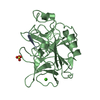
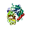
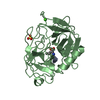
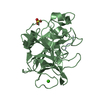
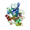
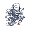
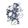
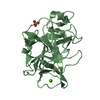
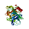
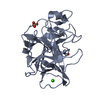
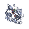
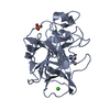
- PDB-2bd7: Porcine pancreatic elastase complexed with beta-casomorphin-7 and... -
+ Open data

- Basic information
| Entry | Database: PDB / ID: 2bd7 | |||||||||
|---|---|---|---|---|---|---|---|---|---|---|
| Title | Porcine pancreatic elastase complexed with beta-casomorphin-7 and Arg-Phe at pH 5.0 (50 min soak) | |||||||||
 Components Components |
| |||||||||
 Keywords Keywords | HYDROLASE / SERINE PROTEINASE | |||||||||
| Function / homology |  Function and homology information Function and homology informationpancreatic elastase / serine-type endopeptidase activity / proteolysis / : / metal ion binding Similarity search - Function | |||||||||
| Biological species |   Homo sapiens (human) Homo sapiens (human) | |||||||||
| Method |  X-RAY DIFFRACTION / MOLECULAR REPLACEMENT / Resolution: 1.6 Å X-RAY DIFFRACTION / MOLECULAR REPLACEMENT / Resolution: 1.6 Å | |||||||||
 Authors Authors | Liu, B. / Schofield, C.J. / Wilmouth, R.C. | |||||||||
 Citation Citation | Journal: J.Biol.Chem. / Year: 2006 Title: Structural analyses on intermediates in serine protease catalysis Authors: Liu, B. / Schofield, C.J. / Wilmouth, R.C. #1: Journal: Nat.Struct.Biol. / Year: 1997Title: Structure of a specific acyl-enzyme complex formed between beta-casomorphin-7 and porcine pancreatic elastase Authors: Wilmouth, R.C. / Clifton, I.J. / Robinson, C.V. / Roach, P.L. / Aplin, R.T. / Westwood, N.J. / Hajdu, J. / Schofield, C.J. #2: Journal: Nat.Struct.Biol. / Year: 2001Title: X-ray snapshots of serine protease catalysis reveal a tetrahedral intermediate Authors: Wilmouth, R.C. / Edman, K. / Neutze, R. / Wright, P.A. / Clifton, I.J. / Schneider, T.R. / Schofield, C.J. / Hajdu, J. | |||||||||
| History |
|
- Structure visualization
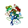
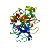
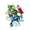
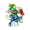
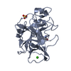
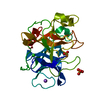
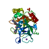
| Structure viewer | Molecule: MolmilJmol/JSmol |
|---|
- Downloads & links
-Download
| PDBx/mmCIF format | 2bd7.cif.gz | 65.4 KB | Display | PDBx/mmCIF format |
|---|---|---|---|---|
| PDB format | pdb2bd7.ent.gz | 46 KB | Display | PDB format |
| PDBx/mmJSON format | 2bd7.json.gz | Tree view | PDBx/mmJSON format | |
| Others |  Other downloads Other downloads |
-Validation report
| Arichive directory | https://data.pdbj.org/pub/pdb/validation_reports/bd/2bd7ftp://data.pdbj.org/pub/pdb/validation_reports/bd/2bd7 | HTTPS FTP |
|---|
-Related structure data
| Related structure data | 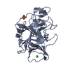 2bb4C  2bd2C 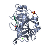 2bd3C  2bd4C  2bd5C 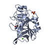 2bd8C 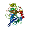 2bd9C 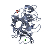 2bdaC 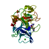 2bdbC 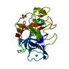 2bdcC  2h1uC 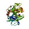 3estS  2bd6 C: citing same article ( S: Starting model for refinement |
|---|---|
| Similar structure data |


-Links
 PDBj
PDBj
- Assembly
| Deposited unit | 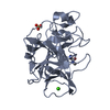
| ||||||||
|---|---|---|---|---|---|---|---|---|---|
| 1 |
| ||||||||
| Unit cell |
|
-Components
| #1: Protein/peptide | Mass: 1168.362 Da / Num. of mol.: 1 / Source method: obtained synthetically / Details: synthetic peptide / Source: (synth.) Homo sapiens (human) | ||
|---|---|---|---|
| #2: Protein | Mass: 25928.031 Da / Num. of mol.: 1 / Source method: isolated from a natural source / Source: (natural) | ||
| #3: Chemical | ChemComp-CA /   Mass: 40.078 Da / Num. of mol.: 1 / Source method: obtained synthetically / Formula: Ca Mass: 40.078 Da / Num. of mol.: 1 / Source method: obtained synthetically / Formula: Ca | ||
| #4: Chemical | ChemComp-SO4 /   Mass: 96.063 Da / Num. of mol.: 1 / Source method: obtained synthetically / Formula: SO4 Mass: 96.063 Da / Num. of mol.: 1 / Source method: obtained synthetically / Formula: SO4 | ||
| #5: Water | ChemComp-HOH /  Mass: 18.015 Da / Num. of mol.: 128 / Source method: isolated from a natural source / Formula: H2O Mass: 18.015 Da / Num. of mol.: 128 / Source method: isolated from a natural source / Formula: H2O | ||
| Has protein modification | Y | ||
| Nonpolymer details | OG SER A 195, C ILE P 7 AND N ARG P 1001 ARE COVALENTLY| Sequence details | THIS CONFLICT IS BASED ON THE REFERENCE 3 IN SEQUENCE DATABASE, P00772 IN SWISS-PROT. | |
-Experimental details
-Experiment
| Experiment | Method: X-RAY DIFFRACTION / Number of used crystals: 1 |
|---|
- Sample preparation
| Crystal | Density Matthews: 2 Å3/Da / Density % sol: 38.59 % |
|---|---|
| Crystal grow | Temperature: 294 K / Method: vapor diffusion, hanging drop / pH: 5 Details: 25MM SODIUM SULPHATE, 25MM SODIUM ACETATE (PH 5.0), 25 MG/ML PPE AND 17.5 MG/ML BCM7, AND THEN SOAKED WITH ARG-PHE (2 MG) IN 250MM SODIUM ACETATE (PH 5.0) (5 MICROLITRES) FOR 50 MIN, VAPOR ...Details: 25MM SODIUM SULPHATE, 25MM SODIUM ACETATE (PH 5.0), 25 MG/ML PPE AND 17.5 MG/ML BCM7, AND THEN SOAKED WITH ARG-PHE (2 MG) IN 250MM SODIUM ACETATE (PH 5.0) (5 MICROLITRES) FOR 50 MIN, VAPOR DIFFUSION, HANGING DROP, temperature 294K |
-Data collection
| Diffraction | Mean temperature: 127 K |
|---|---|
| Diffraction source | Source: ROTATING ANODE / Type: RIGAKU MICROMAX-007 / Wavelength: 1.5418 Å |
| Detector | Type: RIGAKU RAXIS IV / Detector: IMAGE PLATE / Date: Jul 2, 2005 / Details: OSMIC mirrors |
| Radiation | Monochromator: OSMIC MIRRORS / Protocol: SINGLE WAVELENGTH / Monochromatic (M) / Laue (L): M / Scattering type: x-ray |
| Radiation wavelength | Wavelength: 1.5418 Å / Relative weight: 1 |
| Reflection | Resolution: 1.6→19.5 Å / Num. obs: 29030 / % possible obs: 99.8 % / Observed criterion σ(F): 0 / Observed criterion σ(I): 0 / Redundancy: 3.4 % / Biso Wilson estimate: 16.6 Å2 / Rmerge(I) obs: 0.062 / Net I/σ(I): 16.8 |
| Reflection shell | Resolution: 1.6→1.69 Å / Redundancy: 3.3 % / Rmerge(I) obs: 0.204 / Mean I/σ(I) obs: 4 / Num. unique all: 4151 / % possible all: 99.5 |
- Processing
| Software |
| |||||||||||||||||||||||||||||||||||||||||||||||||||||||||||||||||||||||||||||||||||||||||||||||
|---|---|---|---|---|---|---|---|---|---|---|---|---|---|---|---|---|---|---|---|---|---|---|---|---|---|---|---|---|---|---|---|---|---|---|---|---|---|---|---|---|---|---|---|---|---|---|---|---|---|---|---|---|---|---|---|---|---|---|---|---|---|---|---|---|---|---|---|---|---|---|---|---|---|---|---|---|---|---|---|---|---|---|---|---|---|---|---|---|---|---|---|---|---|---|---|---|
| Refinement | Method to determine structure: MOLECULAR REPLACEMENT Starting model: 3EST Resolution: 1.6→19.5 Å / Cor.coef. Fo:Fc: 0.956 / Cor.coef. Fo:Fc free: 0.939 / SU B: 3.191 / SU ML: 0.059 / TLS residual ADP flag: LIKELY RESIDUAL / Cross valid method: THROUGHOUT / σ(F): 0 / ESU R: 0.091 / ESU R Free: 0.091 / Stereochemistry target values: MAXIMUM LIKELIHOOD
| |||||||||||||||||||||||||||||||||||||||||||||||||||||||||||||||||||||||||||||||||||||||||||||||
| Solvent computation | Ion probe radii: 0.8 Å / Shrinkage radii: 0.8 Å / VDW probe radii: 1.2 Å / Solvent model: BABINET MODEL WITH MASK | |||||||||||||||||||||||||||||||||||||||||||||||||||||||||||||||||||||||||||||||||||||||||||||||
| Displacement parameters | Biso mean: 25.487 Å2
| |||||||||||||||||||||||||||||||||||||||||||||||||||||||||||||||||||||||||||||||||||||||||||||||
| Refinement step | Cycle: LAST / Resolution: 1.6→19.5 Å
| |||||||||||||||||||||||||||||||||||||||||||||||||||||||||||||||||||||||||||||||||||||||||||||||
| Refine LS restraints |
| |||||||||||||||||||||||||||||||||||||||||||||||||||||||||||||||||||||||||||||||||||||||||||||||
| LS refinement shell | Resolution: 1.6→1.642 Å / Total num. of bins used: 20
| |||||||||||||||||||||||||||||||||||||||||||||||||||||||||||||||||||||||||||||||||||||||||||||||
| Refinement TLS params. | Method: refined / Origin x: -4.191 Å / Origin y: 28.152 Å / Origin z: 42.862 Å
| |||||||||||||||||||||||||||||||||||||||||||||||||||||||||||||||||||||||||||||||||||||||||||||||
| Refinement TLS group | Selection: ALL |
